Synaptic transmission is the process by which one neuron communicates with another neuron, muscle cell, or gland cell across a specialized junction called a synapse. It is one of the fundamental mechanisms of nervous-system function. Action potentials allow signals to travel along axons, but synaptic transmission allows those signals to influence other cells. Without synapses, neurons would fire in isolation. With synapses, the nervous system becomes a connected network capable of sensation, movement, reflexes, memory, emotion, learning, attention, and thought.
Most synaptic transmission in the nervous system is chemical. When an electrical impulse reaches the end of a neuron’s axon, it triggers the release of neurotransmitters from small vesicles into the synaptic cleft. These neurotransmitters cross the gap and bind to receptors on the next cell, changing its activity. A StatPearls overview describes synaptic transmission as involving neurotransmitter release into the synaptic cleft followed by receptor binding on the postsynaptic cell. The synapse is therefore a place where electrical signaling becomes chemical signaling and then, often, electrical signaling again.
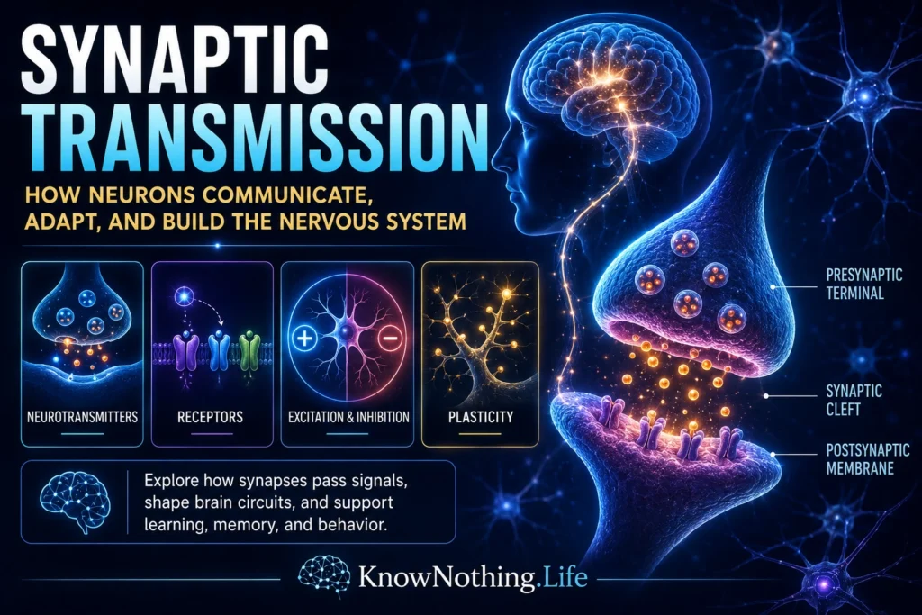
The Structure of a Synapse
A typical chemical synapse has three main parts: the presynaptic terminal, the synaptic cleft, and the postsynaptic membrane. The presynaptic terminal is the end of the sending neuron’s axon. It contains synaptic vesicles filled with neurotransmitter, voltage-gated calcium channels, mitochondria, and specialized protein machinery for vesicle docking and release. The synaptic cleft is the tiny extracellular gap between cells. The postsynaptic membrane contains receptors that detect neurotransmitters and convert chemical signals into cellular responses.
This design allows synapses to be both precise and flexible. The presynaptic neuron controls when neurotransmitter is released. The postsynaptic cell controls how strongly it responds, depending on receptor type, receptor number, membrane voltage, and past activity. Synapses can also be modified by neuromodulators, glial cells, local chemistry, and repeated use. They are not static switches. They are adjustable communication points that can strengthen, weaken, recover, fatigue, adapt, and participate in long-term learning.
From Action Potential to Neurotransmitter Release
Synaptic transmission usually begins when an action potential arrives at the presynaptic terminal. This electrical signal depolarizes the terminal membrane and opens voltage-gated calcium channels. Calcium ions rush into the terminal, creating a local calcium signal near the active zone. Calcium then triggers synaptic vesicles to fuse with the presynaptic membrane, releasing neurotransmitter into the synaptic cleft. Thomas Südhof’s review of calcium control of neurotransmitter release describes this sequence clearly: an action potential opens calcium channels, calcium enters the nerve terminal, and calcium triggers synaptic vesicle exocytosis.
This calcium step is crucial because it links electrical activity to chemical release. The neuron does not simply leak neurotransmitter whenever it is active. It uses highly regulated molecular machinery. Voltage-gated calcium channels help translate the arriving electrical signal into a localized calcium pulse, and that pulse initiates vesicle fusion. A review by Annette Dolphin describes presynaptic calcium channels as the proteins that transduce electrical activity into calcium entry, initiating vesicular neurotransmitter release. This is one of the nervous system’s most elegant transformations: voltage becomes calcium entry, calcium becomes vesicle fusion, and vesicle fusion becomes communication.
Vesicles, SNARE Proteins, and Molecular Machinery
Synaptic vesicles do not release neurotransmitter randomly. They are moved, docked, primed, and fused through specialized molecular machinery. SNARE proteins help bring vesicle and membrane surfaces together so fusion can occur. Synaptotagmins act as calcium sensors, helping link calcium entry to fast vesicle fusion. Complexins, Munc18, Munc13, and other proteins also participate in organizing the release process. A review by Reinhard Jahn explains that calcium-dependent exocytosis of synaptic vesicles is mediated by molecular machines including SNAREs, synaptotagmins, complexins, Munc18, and Munc13.
This machinery matters because synaptic timing must be extraordinarily precise. Speech, vision, balance, reflexes, pain, and movement all depend on neurons communicating within milliseconds. A synapse must release neurotransmitter quickly enough to preserve timing, but also regulate release carefully enough to avoid noise and waste. The molecular complexity of synaptic transmission shows that the nervous system is not just electrical. It is electrochemical, molecular, and highly organized at the smallest scales.
Bernard Katz and Quantal Release
One of the great discoveries in synaptic physiology was quantal release. Bernard Katz and colleagues showed that neurotransmitter is released in small packets, or quanta, corresponding to vesicular release events. His work on the neuromuscular junction helped establish that acetylcholine is stored in vesicles and released in defined amounts. The Nobel Prize summary of Katz’s work notes that he showed acetylcholine is released in highly defined amounts and that calcium plays a key role in triggering this quantal release.
This discovery changed neuroscience because it revealed that synaptic transmission is probabilistic and modular rather than a smooth continuous flow. A presynaptic terminal may release one vesicle, many vesicles, or none in response to activity, depending on calcium entry, vesicle availability, release probability, and synaptic state. Augustine’s historical review of Katz’s work describes how Fatt and Katz concluded that miniature synaptic events arise from spontaneous acetylcholine release from the presynaptic motor neuron. This helped show that even tiny synaptic events could reveal the basic unit structure of neural communication.
Neurotransmitters and Receptors
Neurotransmitters are chemical messengers released at synapses. Major neurotransmitters include glutamate, GABA, glycine, acetylcholine, dopamine, serotonin, norepinephrine, histamine, and many neuropeptides. Glutamate is the principal excitatory neurotransmitter in the brain, while GABA and glycine are major inhibitory neurotransmitters. A StatPearls review of neurotransmitters identifies glutamate as the brain’s principal excitatory neurotransmitter and GABA and glycine as major inhibitory neurotransmitters.
Receptors determine what neurotransmitters do. Ionotropic receptors are ligand-gated ion channels that produce fast electrical effects when neurotransmitters bind. Metabotropic receptors act through G proteins and intracellular signaling pathways, often producing slower and longer-lasting changes. This means the same neurotransmitter can have different effects depending on receptor type and circuit context. Acetylcholine can affect muscle contraction at the neuromuscular junction, attention in the brain, and autonomic function in the body. Dopamine can influence movement, reward, motivation, learning, and decision-making depending on where it acts. Synaptic meaning depends on pathway, receptor, timing, and network state.
Excitation and Inhibition
The nervous system depends on the balance between excitation and inhibition. Excitatory synapses make the postsynaptic cell more likely to fire by depolarizing its membrane. Inhibitory synapses make firing less likely by hyperpolarizing the membrane or stabilizing it away from threshold. Glutamate often produces excitatory postsynaptic potentials, while GABA and glycine commonly produce inhibitory effects. A StatPearls review of GABA receptors describes GABA as reducing neuronal excitability by causing hyperpolarization and decreasing the likelihood of firing.
This balance is not merely technical; it is essential to brain function. Too little excitation can prevent circuits from activating. Too little inhibition can produce runaway activity, instability, or seizures. Attention, movement, perception, and thought all require the brain to amplify some signals while suppressing others. Inhibition is not the opposite of function. It is one of the main ways function becomes precise. A healthy nervous system depends on controlled activity, not maximum activity.
Synaptic Integration
A single neuron may receive thousands of synaptic inputs across its dendrites and cell body. Some inputs are excitatory, some inhibitory, some fast, some slow, and some modulatory. Synaptic integration is the process by which the neuron combines these signals and determines whether to fire an action potential. Inputs arriving close together in time can summate. Inputs arriving on different parts of the dendritic tree can interact. Inhibitory inputs can cancel or shape excitatory ones. The result is not a simple yes-or-no response to one signal, but a dynamic calculation across many inputs.
This is one reason neurons are more than wires. Dendrites can perform local computations, receptors can change sensitivity, and synapses can be strengthened or weakened by prior activity. A neuron’s output reflects its history, its current inputs, and the state of the larger circuit. Synaptic transmission is therefore the cellular foundation of information processing. It allows the nervous system to compare, filter, select, combine, and transform signals.
Synaptic Plasticity and Learning
Synapses can change with experience. This property, called synaptic plasticity, is one of the major biological foundations of learning and memory. Some forms of plasticity last milliseconds or seconds, allowing synapses to adapt during ongoing activity. Other forms last much longer. Long-term potentiation, or LTP, is a long-lasting increase in synaptic strength after repeated activity. A review by Robert Nicoll describes LTP, discovered by Bliss and Lømo in 1973, as a compelling cellular model for learning and memory.
Long-term depression, or LTD, is a long-lasting weakening of synaptic strength. Together, potentiation and depression allow circuits to be refined by experience. Learning does not require every synapse to become stronger. It requires patterns of connection to become better organized. Some pathways are reinforced, others are weakened, and networks become more efficient. This is how practice can change skill, memory can change behavior, and repeated experience can reshape the nervous system.
Synapses, Glial Cells, and the Wider Neural Environment
Synaptic transmission is often described as communication between two neurons, but the surrounding environment also matters. Astrocytes help regulate extracellular ions, remove neurotransmitters, influence metabolism, and participate in synaptic function. Microglia help shape synaptic pruning and immune surveillance. Oligodendrocytes influence conduction through myelin. The old view of glial cells as passive support cells has been replaced by a more active model in which glia help maintain and regulate neural communication.
This wider environment is important because synapses are chemically sensitive. Neurotransmitter must be released, detected, cleared, recycled, and regulated. Too much neurotransmitter in the synaptic cleft can distort signaling. Too little can weaken communication. Transporters, enzymes, receptors, vesicle recycling, and glial uptake all help maintain synaptic balance. Communication in the nervous system is not just about sending a signal. It is about sending the right signal, at the right time, in the right amount, and then clearing the signal so the next one can be understood.
Clinical Importance of Synaptic Transmission
Many neurological and psychiatric conditions involve synaptic transmission. Epilepsy can involve abnormal excitation and inhibition. Depression, anxiety, schizophrenia, Parkinson’s disease, Alzheimer’s disease, addiction, migraine, and many movement disorders involve neurotransmitter systems, synaptic plasticity, receptor function, or circuit regulation. Many medications act at synapses. Antidepressants can influence serotonin or norepinephrine signaling. Antipsychotics often affect dopamine receptors. Benzodiazepines enhance GABA-related inhibition. Stimulants affect dopamine and norepinephrine. Anesthetics, anticonvulsants, pain medications, and many toxins also alter synaptic function.
Synaptic dysfunction can also occur in neurodegenerative disease. Before large-scale cell death appears, synaptic changes may already impair communication. This is especially important in disorders affecting memory, cognition, and movement. The brain does not need to lose every neuron for function to decline; it can lose the coordination and strength of communication among neurons. Synapses are therefore central to both normal function and disease.
Why Synaptic Transmission Matters
Synaptic transmission matters because it is the process that turns individual neurons into a nervous system. Action potentials carry signals within neurons, but synapses allow signals to move between cells, change circuits, influence behavior, and adapt through learning. Every sensation, movement, memory, emotion, reflex, and thought depends on synaptic communication.
The deeper lesson is that the mind depends on connection. A neuron alone can fire, but networks require synapses. Synaptic transmission allows the brain to be flexible, selective, and plastic. It lets experience shape the nervous system and lets the nervous system shape behavior. To understand synaptic transmission is to understand one of neuroscience’s central truths: the brain works not only because neurons exist, but because they communicate, adapt, and form living circuits.