
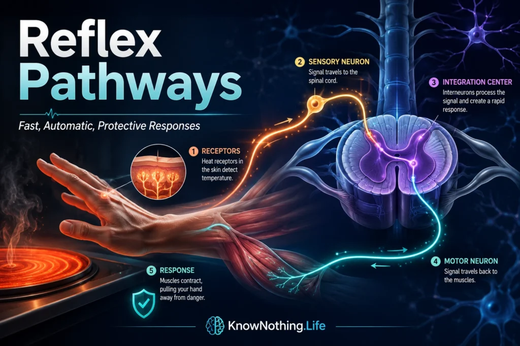
Reflex pathways are neural circuits that produce rapid, automatic responses to specific stimuli. They allow the body to react before conscious thought has fully analyzed what is happening. When a hand pulls away from heat, a knee jerks after a tendon tap, pupils change size in response to light, or posture adjusts after a stumble, reflex pathways are at work. These responses are not random twitches. They are organized patterns of sensory input, neural processing, and motor output that help protect the body, maintain posture, regulate organs, and support survival.
A reflex pathway is often described through the reflex arc, the basic unit of reflex action. In a typical reflex arc, a receptor detects a stimulus, a sensory neuron carries information toward the spinal cord or brainstem, an integration center processes the signal, a motor neuron carries the response outward, and an effector such as a muscle or gland produces the action. StatPearls describes the reflex arc as the basic unit of a reflex and emphasizes that reflex pathways can act on an impulse before that impulse reaches full conscious processing in the brain.
The Reflex Arc: Receptor, Sensory Neuron, Integration, Motor Neuron, Effector
The classic reflex arc begins with a receptor. This receptor may be a muscle spindle detecting stretch, a nociceptor detecting damaging heat, a photoreceptor-related pathway responding to light, or a visceral receptor monitoring internal organ state. Once the stimulus is detected, sensory or afferent neurons carry the signal into the central nervous system. Some reflexes are processed mainly in the spinal cord, while others involve the brainstem or autonomic circuits. The response then leaves through motor or efferent pathways, producing contraction, withdrawal, gland secretion, pupil change, cardiovascular adjustment, or another automatic response.
Reflexes are fast because they reduce the number of steps required before action. A spinal reflex does not need to wait for detailed cortical interpretation before protecting the body. That does not mean the brain is uninvolved altogether. The brain may receive sensory information shortly afterward, interpret the event, remember it, and adjust future behavior. Reflex pathways therefore work at two levels: immediate protection and later conscious understanding. They give the nervous system a way to act now and analyze later.
Monosynaptic Reflexes and the Stretch Reflex
The simplest reflexes are monosynaptic reflexes, meaning the sensory neuron communicates directly with the motor neuron through a single synapse. The best-known example is the stretch reflex, also called the muscle stretch reflex or deep tendon reflex. When a tendon is tapped, it briefly stretches the muscle. Muscle spindles detect that stretch and send signals through Ia afferent fibers into the spinal cord. These fibers synapse directly onto alpha motor neurons, which activate the same muscle and cause it to contract. StatPearls describes the monosynaptic stretch reflex as a reflex arc that allows direct communication between sensory and motor neurons innervating the muscle.
The stretch reflex is important for posture and muscle tone. If a muscle is unexpectedly stretched, the reflex helps resist that stretch and stabilize the body. This is why deep tendon reflexes are useful in neurological examinations. A reflex hammer does not merely test whether a muscle can move. It tests the integrity of sensory fibers, spinal segments, motor neurons, neuromuscular junctions, and descending influences from the brain. StatPearls describes the myotatic reflex as involving structures such as the muscle spindle, primary afferent sensory neuron, inhibitory interneuron, alpha motor neuron, and gamma motor neuron.
Polysynaptic Reflexes and Withdrawal
Many reflexes are polysynaptic, meaning one or more interneurons are involved between the sensory and motor neurons. The withdrawal reflex is a classic example. If a person touches something sharp or hot, nociceptors send signals into the spinal cord. Interneurons then activate flexor muscles that pull the affected limb away from the harmful stimulus. This can happen before the person has fully identified what caused the pain. StatPearls describes the withdrawal response as a protective reflex that removes the body from potentially damaging stimuli.
The withdrawal reflex is more complex than a simple muscle contraction. It may require activating some muscles while inhibiting others. If the foot steps on something painful, the leg must flex to withdraw, while the opposite leg may extend to support body weight. This is called the crossed extension reflex. NCBI’s Neuroscience text describes the crossed extension reflex as enhancing postural support during withdrawal of the affected limb from a painful stimulus. This shows that even “simple” reflexes are coordinated actions. They protect one body part while preserving balance and whole-body stability.
Reflexes, Inhibition, and Coordination
Reflex pathways depend heavily on inhibition. If a muscle contracts, its antagonist often needs to relax so the movement can happen smoothly. This is called reciprocal inhibition. For example, when the stretch reflex activates one muscle, inhibitory interneurons may reduce activation in the opposing muscle. Without this coordination, the body would fight itself, producing stiff, inefficient, or unstable movement. Reflexes therefore depend not only on activation, but also on carefully timed restraint.
This principle connects reflex pathways to larger motor control. The spinal cord is not a passive cable carrying commands from the brain to muscles. It contains interneuron networks that integrate sensory input, shape motor output, coordinate reflexes, and interact with descending pathways from the brain. Lower motor neurons also participate in somatic reflex arcs, either through direct monosynaptic connections or through polysynaptic pathways involving interneurons. Reflexes are therefore part of the nervous system’s layered design: local circuits handle urgent control, while higher centers modulate them according to context.
Sherrington and the Integrative Action of the Nervous System
The scientific understanding of reflex pathways was deeply shaped by Charles Scott Sherrington. His 1906 book The Integrative Action of the Nervous System helped establish reflexes as basic units of nervous-system organization. Sherrington argued that the reflex was the simplest unit of nervous integration and introduced the synapse as the site where elementary reflexes interact to produce more complex, unified behavior.
Sherrington’s importance lies in the word “integration.” Reflexes are not isolated mechanical reactions. They are coordinated within the larger organism. A withdrawal reflex must fit posture, balance, muscle tone, and ongoing movement. A postural reflex must stabilize the body without preventing voluntary motion. A reflex pathway therefore shows how the nervous system combines local automatic responses into whole-body behavior. Sherrington recognized that the spinal cord, though simpler than the cerebrum, displays core principles of synaptic nervous-system function.
Brainstem and Autonomic Reflexes
Not all reflex pathways are spinal. Many involve the brainstem, especially reflexes related to survival, cranial nerves, and autonomic control. The pupillary light reflex adjusts pupil size in response to light. The corneal reflex helps protect the eye. The gag reflex protects the airway. Coughing, sneezing, swallowing, vomiting, and vestibulo-ocular reflexes all involve brainstem circuits. These reflexes help regulate breathing, feeding, vision, balance, airway protection, and sensory-motor coordination.
Autonomic reflexes regulate internal organs. Baroreceptor reflexes help maintain blood pressure. Digestive reflexes coordinate gut movement and secretion. Bladder reflexes help regulate urination. These pathways often involve the autonomic nervous system, including sympathetic and parasympathetic branches. Like somatic reflexes, autonomic reflexes use sensory information to produce an adaptive response. The difference is that the effectors are often smooth muscle, cardiac muscle, or glands rather than skeletal muscles.
Reflexes and Voluntary Movement
Reflexes are often contrasted with voluntary movement, but the two are deeply connected. Voluntary movement depends on reflex pathways for stability, feedback, and correction. When someone walks, spinal reflexes help adjust muscle tone and respond to changes in surface or load. When someone lifts an object heavier than expected, stretch and load-related reflexes help correct grip and posture. When a person stumbles, rapid postural reflexes help prevent a fall before conscious planning can intervene.
The brain can also modulate reflexes. Anxiety, attention, fatigue, injury, training, medication, and neurological disease can all change reflex strength. Athletes and musicians do not eliminate reflexes; they train motor systems so reflexes, sensory feedback, and voluntary commands work together more efficiently. Reflex pathways therefore are not primitive leftovers beneath intelligence. They are essential components of skilled action.
Clinical Importance of Reflex Pathways
Reflex testing is a major part of neurological examination because reflexes reveal the integrity of specific pathways. Reduced or absent reflexes may suggest problems in peripheral nerves, sensory fibers, motor neurons, nerve roots, or muscles. Exaggerated reflexes may suggest upper motor neuron involvement, where descending control from the brain or spinal cord has been disrupted. Asymmetry between the two sides of the body can help localize neurological damage. The deep tendon reflex is therefore a small clinical test with large diagnostic value.
Reflex abnormalities can appear in stroke, spinal cord injury, neuropathy, radiculopathy, motor neuron disease, multiple sclerosis, traumatic brain injury, metabolic disorders, and many other conditions. Reflexes are useful because they test circuits rather than isolated anatomy. A tendon tap tests receptor function, sensory conduction, spinal integration, motor output, neuromuscular transmission, and muscle response in one brief event. This is why physicians can learn so much from a simple reflex hammer.
Why Reflex Pathways Matter
Reflex pathways matter because they show that the nervous system is built for fast, adaptive action. The body cannot wait for slow conscious analysis every time it encounters heat, pain, imbalance, bright light, airway irritation, or sudden stretch. Reflexes provide immediate responses that protect tissue, preserve posture, regulate organs, and support survival. They are automatic, but they are not crude. They are organized circuits shaped by anatomy, physiology, inhibition, feedback, and integration.
The deeper lesson is that intelligence begins in action. The nervous system does not only think about the world; it must respond to it. Reflex pathways reveal how sensation becomes movement, how danger becomes protection, and how the body maintains control beneath awareness. To understand reflex pathways is to understand one of neuroscience’s most basic principles: before the brain reflects, the nervous system reacts, stabilizes, protects, and keeps the organism alive.


