
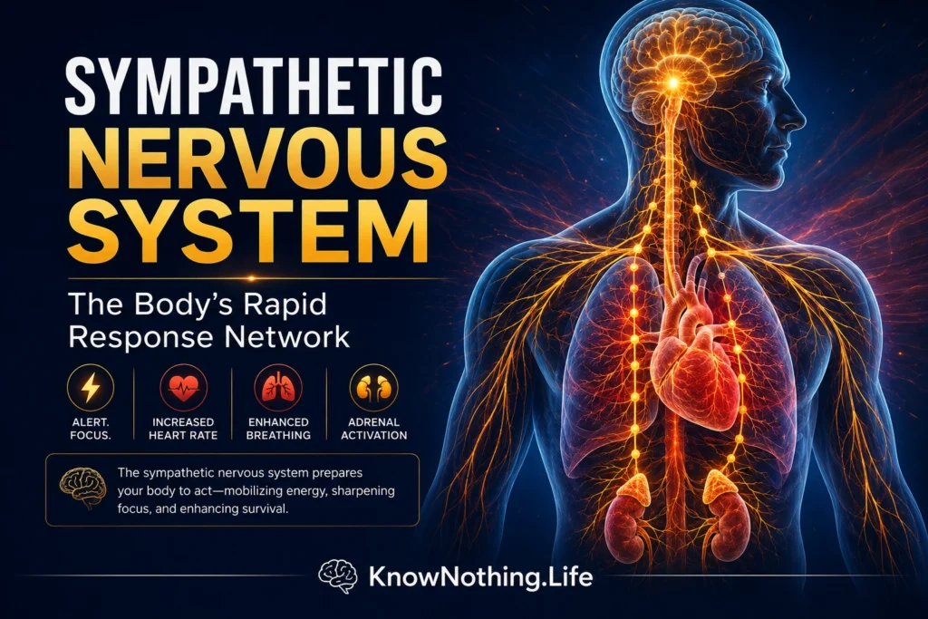
The sympathetic nervous system is one division of the autonomic nervous system, the network that regulates internal organs, blood vessels, glands, and metabolism without requiring continuous conscious control. It is often described as the body’s “fight-or-flight” system, a phrase associated with Walter Cannon’s early twentieth-century work on emergency physiology. In Bodily Changes in Pain, Hunger, Fear and Rage, Cannon described how threats produce coordinated changes in circulation, breathing, digestion, and energy availability. Yet the sympathetic system is not reserved for crises. It remains active throughout ordinary life, adjusting blood pressure when a person stands, redistributing blood flow during exercise, regulating sweating and skin temperature, and supporting circulation during sleep, illness, and environmental change.
Modern research has replaced the idea of a single on-or-off alarm with a model of patterned autonomic control. Sympathetic outflow can increase to one organ while remaining stable or decreasing elsewhere, allowing the body to respond selectively. Michael Esler and colleagues demonstrated this regional organization by measuring organ-specific norepinephrine spillover in humans. Their findings showed distinct contributions from the kidneys, skeletal muscles, lungs, skin, heart, and abdominal organs, illustrating why a blood measurement of total norepinephrine cannot fully represent sympathetic activity throughout the body. The system is therefore best understood as a distributed network that produces responses matched to particular physiological demands.
Anatomy and Chemical Communication
Sympathetic pathways begin with preganglionic neurons located mainly in the thoracic and upper lumbar spinal cord. Their axons leave the spinal cord and travel to sympathetic ganglia positioned beside or in front of the vertebral column. There they communicate with postganglionic neurons whose axons extend to blood vessels, the heart, sweat glands, digestive organs, kidneys, pupils, and other targets. Some preganglionic fibers pass directly to the adrenal medulla, where they stimulate chromaffin cells to release catecholamines into the bloodstream. This arrangement gives sympathetic control both a fast neural component and a broader hormonal component.
Acetylcholine is released by sympathetic preganglionic neurons, while most postganglionic sympathetic terminals release norepinephrine. Important exceptions include sympathetic fibers supplying sweat glands, which also use acetylcholine. Ulf von Euler’s work identifying norepinephrine as the principal transmitter of many sympathetic nerve endings was foundational to autonomic pharmacology. Norepinephrine acts through alpha- and beta-adrenergic receptors, and its effects depend on receptor type and tissue. In blood vessels it commonly promotes constriction through alpha receptors, while cardiac beta-1 receptors increase heart rate and contractile strength. Most released norepinephrine is taken back into nerve terminals rather than entering the circulation, a recycling process documented through cardiac norepinephrine and metabolite measurements.
Cardiovascular Control and the Baroreflex
One of the sympathetic system’s most important responsibilities is maintaining adequate blood pressure and organ perfusion. When a person stands, gravity shifts blood toward the legs and abdomen. Pressure-sensitive baroreceptors in the carotid arteries and aortic arch detect the reduction in arterial stretch and relay information to the brainstem. Sympathetic activity then increases to the heart and blood vessels, accelerating the heart, strengthening contraction, and narrowing selected vessels to preserve blood flow to the brain. When pressure rises, baroreflex pathways suppress sympathetic vasoconstrictor activity. This negative-feedback system operates from heartbeat to heartbeat rather than waiting for a conscious perception of dizziness.
The baroreflex also illustrates why sympathetic regulation is dynamic rather than fixed. Exercise, hypoxia, emotion, and disease can shift the operating point of the reflex while preserving its capacity to respond to rapid pressure changes. Direct recordings of muscle sympathetic nerve activity show that bursts are closely related to fluctuations in arterial pressure. In people with postural orthostatic tachycardia syndrome, Norman Swift and colleagues found exaggerated sympathetic responses to baroreflex challenges even though resting activity did not necessarily differ from healthy controls. Autonomic symptoms can therefore arise from abnormal reflex responsiveness rather than simply from continuously elevated sympathetic tone.
Exercise, Stress, and Immediate Adaptation
During physical activity, the sympathetic nervous system helps match circulation to metabolic demand. Heart rate and cardiac output rise, blood flow is redirected, and vascular resistance is adjusted so active muscles receive sufficient oxygen without causing arterial pressure to collapse. Signals arise from central motor command as the brain initiates movement, from pressure-sensitive receptors, and from sensory fibers in contracting muscles. Studies using sustained handgrip have shown that muscle sympathetic nerve activity rises as exercise becomes more intense and fatiguing. Robert Victor and Douglas Seals also demonstrated that chemical signals produced in working muscle contribute to the exercise pressor reflex, increasing sympathetic outflow to nonworking regions.
Psychological stress can activate sympathetic pathways even when no physical movement occurs. In a human microneurography study, Matthew Muller and colleagues found that mental arithmetic produced rapid and reproducible increases in skin sympathetic nerve activity. This response can alter sweating, skin blood flow, cardiac function, and vascular tone. However, mental stress does not raise all measures of sympathetic activity equally. These observations support a broader interpretation of Cannon’s emergency response: the sympathetic system prepares the body for action through organ-specific patterns shaped by cognition, emotion, posture, temperature, and current physiological need.
Metabolism, Temperature, and Immune Regulation
Sympathetic signaling affects energy use as well as circulation. During demanding conditions, catecholamines promote the availability of glucose and fatty acids, helping supply fuel to the brain, heart, and skeletal muscles. Sympathetic nerves also participate in temperature regulation by controlling skin blood vessels, sweat glands, and thermogenic tissues. Cold exposure generally increases sympathetic drive to conserve heat through vasoconstriction and to produce heat through metabolic mechanisms. Human studies of brown adipose tissue have shown that cold-responsive thermogenesis is associated with sympathetic activation, although systemic stimulation of beta-adrenergic receptors does not always reproduce the same localized brown-fat response.
The sympathetic nervous system also communicates with the immune system. Sympathetic fibers innervate lymphoid organs, and norepinephrine can influence immune-cell migration, cytokine production, and inflammatory activity. The outcome depends on receptor distribution, cell type, disease stage, and the timing of sympathetic signaling. A controlled human experiment by Matthijs Kox and colleagues found that voluntary activation techniques associated with increased epinephrine were followed by reduced production of several proinflammatory mediators after experimental endotoxin administration. The study did not establish that sympathetic activation is universally beneficial, but it showed that autonomic activity can measurably alter innate immune responses in humans.
Chronic Overactivity, Disease, and Clinical Importance
Short-term sympathetic activation is adaptive, but persistent or poorly regulated activation can become damaging. Chronic sympathetic drive increases cardiac workload, promotes vascular constriction, alters kidney function, and may contribute to hypertension, arrhythmias, metabolic dysfunction, and heart failure progression. In a landmark study of patients with congestive heart failure, Gary Hasking and colleagues measured markedly increased total, cardiac, and renal norepinephrine spillover. Later work showed that cardiac sympathetic activation may become especially pronounced, exposing the failing heart to greater adrenergic stimulation. What initially helps preserve blood pressure and cardiac output can therefore become part of a self-reinforcing disease process.
Sympathetic dysfunction can appear as insufficient activation, excessive activation, or inappropriate regional distribution. Patients may experience orthostatic dizziness, abnormal sweating, heat intolerance, palpitations, unstable blood pressure, or exercise intolerance. Clinicians assess the system through blood-pressure responses, autonomic reflex tests, sweat testing, catecholamine measurements, heart-rate analysis, and, in specialized laboratories, microneurography. This technique records postganglionic sympathetic traffic directly from a peripheral nerve and has revealed that sympathetic bursts vary with breathing, pressure, sleep, emotion, and disease. The sympathetic nervous system is therefore neither an enemy to be permanently “calmed” nor a simple stress switch. It is an essential adaptive network whose value depends on precision, timing, and recovery after a challenge has passed.


