
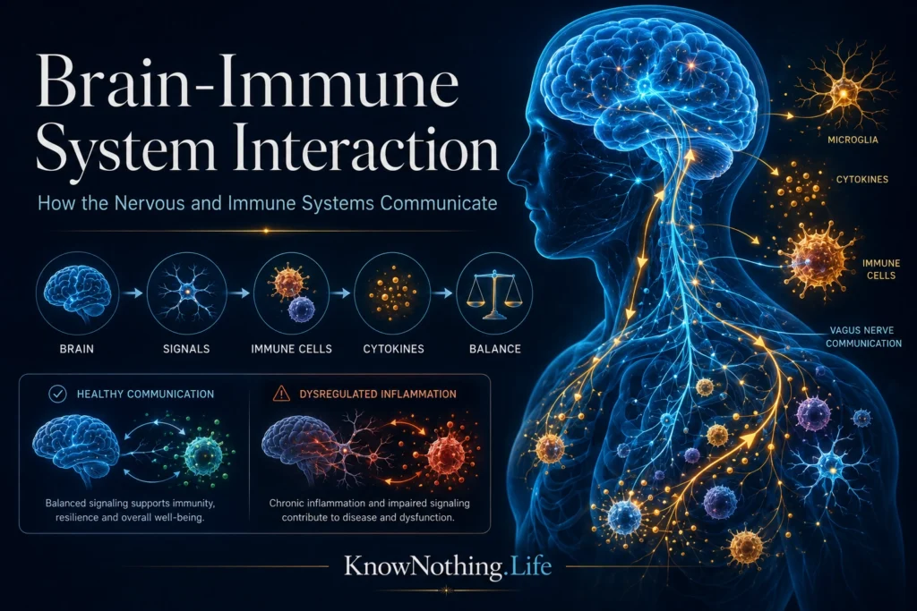
Brain-immune system interaction refers to the continuous exchange of information between the nervous system and the body’s defenses. Immune cells and signaling molecules report infection, injury, metabolic disruption, and tissue stress to the brain. The brain then coordinates fever, appetite, sleep, hormone release, autonomic activity, and behavior while sending signals that can intensify or restrain inflammation. The two systems are therefore not independent command centers. They form an integrated regulatory network designed to preserve stability while responding to danger.
This relationship is essential in health, not merely a feature of disease. Immune-related molecules help shape neural development, synaptic remodeling, tissue repair, and the removal of damaged cells. Neural circuits, in turn, influence the movement and activity of immune cells in organs such as the spleen, bone marrow, gut, and lymph nodes. When communication becomes excessive, prolonged, or misdirected, it can contribute to autoimmune disease, neurodegeneration, chronic pain, mood symptoms, and cognitive changes.
How the Brain Detects Immune Activity
Immune signals reach the brain through several routes. Cytokines released during infection or injury can act on blood vessels and specialized regions where the blood-brain barrier is more accessible to circulating information. Brain endothelial cells translate peripheral inflammatory signals into local chemical messages, while immune activity near the meninges and cerebrospinal fluid provides additional information. The blood-brain barrier is not an absolute wall but a regulated interface whose permeability and signaling properties can change during systemic inflammation.
The discovery of functional lymphatic vessels around the brain further revised the idea that the central nervous system is isolated from conventional immune drainage. In 2015, Antoine Louveau and colleagues identified lymphatic vessels lining the dural sinuses of mice. These structures carried fluid and immune cells from cerebrospinal fluid toward deep cervical lymph nodes, providing a route through which the immune system can monitor substances leaving the central nervous system. The finding did not eliminate the concept of specialized brain immunity, but it revealed a more direct anatomical connection between the brain and peripheral immune organs.
Neural Pathways That Sense Inflammation
Neural routes allow the brain to detect immune activity rapidly. Sensory fibers of the vagus nerve monitor conditions within the heart, lungs, digestive tract, liver, and other organs before transmitting information to the brainstem. In a 2016 study, Benjamin Steinberg and colleagues recorded different patterns of vagal electrical activity after administering specific cytokines. Their results suggested that vagal nerves can encode features of an immune response rather than communicating only a general signal that the body is unwell.
A 2024 study led by Hao Jin provided an even more detailed picture of this communication in mice. The researchers identified distinct populations of vagal sensory neurons that responded to pro-inflammatory and anti-inflammatory cytokines. These signals reached brainstem circuits that could subsequently increase or suppress inflammatory responses in the body. The work revealed a bidirectional system in which the brain receives relatively specific information about peripheral immunity and then adjusts that response through neural output.
How the Brain Regulates Immunity
The brain influences immune activity through endocrine and autonomic pathways. Activation of the hypothalamic-pituitary-adrenal axis releases glucocorticoids, which can limit inflammatory gene expression and prevent immune responses from escalating without control. Sympathetic nerves release catecholamines in lymphoid organs and other tissues, affecting immune-cell movement, cytokine production, blood flow, and energy availability. These responses can be protective during a short-term threat, although repeated activation may produce different effects from a brief, well-regulated stress response.
One of the field’s most influential discoveries was the identification of the inflammatory reflex. In 2000, Linda Borovikova and colleagues found that electrically stimulating the vagus nerve reduced the production of tumor necrosis factor and protected animals during experimental endotoxemia. The study showed that efferent neural activity could suppress systemic inflammation rather than merely detect it. Later research connected this pathway with the splenic nerve and acetylcholine-sensitive immune cells, laying the foundation for bioelectronic medicine—the attempt to treat inflammatory disease by stimulating defined neural circuits.
Microglia and Immunity Inside the Brain
Microglia are the principal resident immune cells of the brain and spinal cord. They constantly survey their surroundings, remove cellular debris, respond to infection, and help coordinate tissue repair. Fate-mapping experiments published by Florent Ginhoux and colleagues in 2010 showed that adult microglia arise largely from primitive embryonic macrophage precursors rather than being routinely replaced by circulating adult monocytes. Their long residence within nervous tissue allows them to respond closely to local neural activity, but it also means that prolonged inflammatory conditions can alter their behavior over time.
Microglia also perform important functions during healthy brain development. Beth Stevens and colleagues reported in 2007 that proteins belonging to the classical complement system help tag selected synapses for elimination. Microglia recognize these signals and remove connections as neural circuits mature. This mechanism is useful during development, but similar processes may become damaging when reactivated during disease. In Alzheimer’s mouse models, Soyon Hong and colleagues found that blocking complement proteins or a microglial complement receptor reduced early synaptic loss, illustrating how a normal remodeling system may contribute to degeneration under abnormal conditions.
Sickness Behavior as a Brain Response
Fatigue, reduced appetite, social withdrawal, sleepiness, and diminished motivation commonly accompany infection. These changes were once treated as inconvenient side effects of illness, but research on sickness behavior shows that they are coordinated responses organized by the brain. Pro-inflammatory cytokines influence hypothalamic and brainstem circuits that redirect energy away from exploration, reproduction, and strenuous activity toward immune defense and recovery. Feeling ill is therefore partly a neural program activated by immune information rather than simply a direct consequence of damaged tissue.
Modern experiments have begun identifying the circuits responsible for individual elements of sickness. In 2022, Jessica Osterhout and colleagues described a population of neurons in the ventral medial preoptic area of the mouse hypothalamus that became active after immune challenges. Manipulating these neurons altered fever, warmth-seeking, and appetite suppression. The findings explain how a peripheral immune event can produce coordinated changes in temperature, motivation, and behavior, while also showing that different sickness symptoms are generated through organized neural pathways.
Stress, Learning, and Immune Function
The brain can influence immunity through learned associations as well as direct physiological pathways. In 1975, Robert Ader and Nicholas Cohen demonstrated conditioned immunosuppression in rats by pairing a sweetened drink with an immunosuppressive drug. After conditioning, the taste alone reproduced part of the immune effect. Later experiments showed that conditioned immune responses could even modify the progression of autoimmune disease in animals. This work helped establish psychoneuroimmunology by demonstrating that learning can affect measurable immune processes rather than merely changing subjective feelings.
Human research has connected prolonged psychological stress with vulnerability to infection. In a controlled viral-challenge study published in 1991, Sheldon Cohen and colleagues found a dose-response relationship between reported stress and the likelihood of developing respiratory infection. Animal research suggests possible mechanisms. Eric Wohleb and colleagues reported that repeated social stress recruited bone-marrow-derived monocytes to the mouse brain and promoted anxiety-like behavior. Such findings do not mean stress inevitably causes disease, but they demonstrate how neural, hormonal, and immune responses can reinforce one another.
The Gut, Microbiota, and Brain Immunity
The digestive system contains an extensive immune network and a dense community of microorganisms, making it an important site of brain-immune communication. Microbial metabolites can enter circulation, influence immune cells, alter intestinal-barrier function, and affect sensory signals traveling through the vagus nerve. Diet, infection, antibiotics, aging, and disease can all change this ecosystem. However, the term gut-brain axis should not be used as a simple explanation for every neurological or psychiatric symptom.
Animal research has nevertheless identified important biological mechanisms. In 2015, David Erny and colleagues found that germ-free mice developed immature and functionally altered microglia. Some abnormalities could be improved through microbial products, including short-chain fatty acids, indicating that signals generated by intestinal microorganisms contribute to normal microglial development and activity. Translation to humans remains difficult because human microbiomes are highly variable and many findings come from tightly controlled mouse models. The evidence supports genuine communication, not claims that one diet or probiotic can reliably treat complex brain disease.
Brain-Immune Interaction in Disease
Disrupted communication can take several forms. In multiple sclerosis, immune activity damages myelin and other structures in the central nervous system. A 2008 randomized trial led by Stephen Hauser showed that depleting B cells with rituximab reduced inflammatory brain lesions and clinical relapses in people with relapsing-remitting multiple sclerosis. The results demonstrated that a defined immune-cell population could influence the course of a neurological disorder and helped change scientific understanding of the disease.
In anti-NMDA receptor encephalitis, antibodies bind to neuronal receptors and cause them to be removed from the cell surface, weakening synaptic communication. Research led by Ethan Hughes showed that patients’ antibodies selectively reduced surface NMDA receptors and NMDA-mediated currents in neurons. Immune mechanisms are also being studied in stroke, traumatic injury, chronic pain, and neurodegenerative disease. Their effects depend on timing: an early response may remove damaged tissue and support repair, while prolonged activation can injure neurons, synapses, or the blood-brain barrier.
The Future of Brain-Immune Research
The future of the field lies in identifying the precise circuits, immune-cell states, and molecular signals operating in individual diseases. Single-cell methods are revealing microglial, macrophage, and lymphocyte populations that were previously grouped together. Improved imaging may make it possible to measure blood-brain barrier disruption, meningeal lymphatic drainage, and neuroinflammation more accurately. Neural stimulation could eventually regulate selected immune pathways without producing the broad effects associated with systemic immunosuppressive drugs.
The central lesson is that the brain does not merely receive immune messages, and the immune system does not operate independently of neural activity, behavior, or experience. Each system continuously senses and adjusts the other. Their dialogue can produce fever, fatigue, learning, healing, and protection from infection, but it can also contribute to chronic disease when regulation fails. Understanding brain-immune system interaction therefore offers a more complete model of health—one in which neurological, psychological, endocrine, and immune processes are interconnected parts of the same adaptive system.


