
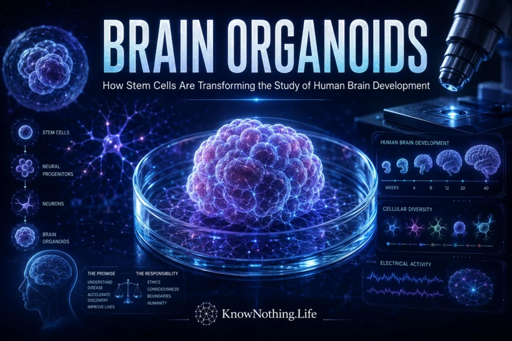
Brain organoids are three-dimensional clusters of human neural tissue grown from pluripotent stem cells. Under controlled laboratory conditions, stem cells multiply, specialize into neural cell types, and organize into structures that reproduce selected features of the developing brain. Depending on the protocol, an organoid may contain ventricular-like zones, neural progenitors, cortical neurons, astrocytes, retinal cells, or cells resembling those of the forebrain and midbrain. They are more biologically complex than flat cell cultures, but they are not complete miniature brains. They lack the full anatomy, sensory systems, blood supply, bodily connections, and developmental history of a human brain.
The modern field was established by Madeline Lancaster and colleagues in 2013. Their cerebral organoids formed recognizable brain-region-like tissues and reproduced features of cortical development. When generated from cells carrying a CDK5RAP2 mutation associated with primary microcephaly, the organoids developed smaller neuroepithelial regions and showed premature neural differentiation. The study demonstrated that a disorder of human brain growth could be reconstructed in patient-derived tissue outside the body.
How Brain Organoids Are Created
Most brain organoids begin with embryonic stem cells or induced pluripotent stem cells. Induced pluripotent stem cells are adult cells, often taken from skin or blood, that have been reprogrammed into a state capable of forming many cell types. Researchers aggregate them into small spheres, expose them to signals that encourage neural development, and frequently embed them in a supportive extracellular matrix. Rotating bioreactors or orbital shakers improve the distribution of oxygen and nutrients while the tissue grows over weeks or months.
Unguided cerebral organoids rely heavily on self-organization and can generate a mixture of regional identities. Guided organoids receive defined molecular signals that push development toward a particular structure, such as the dorsal forebrain, ventral forebrain, midbrain, cerebellum, or choroid plexus. Guided methods generally offer greater experimental control, while unguided systems may reveal unexpected interactions. Neither approach grows a scaled-down adult brain; organoids most closely reproduce selected stages of prenatal development.
Self-Organization and Cellular Diversity
One of the most important properties of organoids is self-organization. Through gene expression, chemical gradients, mechanical forces, and communication with neighboring cells, stem cells can form polarized progenitor zones and generate neurons in developmentally ordered patterns. Giorgia Quadrato and colleagues analyzed more than 80,000 cells from 31 brain organoids in 2017 and found populations related to the cerebral cortex, retina, and other endogenous cell classes. Organoids maintained for more than nine months also developed dendritic spines and spontaneously active neuronal networks.
Reproducibility has nevertheless been a major concern. If two organoids made with the same method contain different cell populations, an apparent disease effect may reflect random variation. Silvia Velasco and colleagues addressed this problem in 2019 using a directed dorsal-forebrain protocol. Single-cell sequencing of more than 166,000 cells showed that 95 percent of the organoids produced a closely similar collection of cortical cell types and followed comparable developmental trajectories. Reproducibility can therefore improve when regional patterning and culture conditions are tightly controlled.
Modeling Developmental Disease and Infection
Brain organoids are particularly useful for studying disorders that begin before birth. Patient-derived induced pluripotent stem cells preserve an individual’s genetic variants, allowing affected organoids to be compared with gene-corrected controls. Researchers can examine whether a mutation changes progenitor proliferation, neuronal migration, cell survival, synapse formation, or the timing of differentiation. Organoids have been applied to microcephaly, epilepsy, autism-associated syndromes, lissencephaly, fragile X syndrome, and other developmental conditions.
The Zika virus epidemic provided an early demonstration of their value. In 2016, Xuyu Qian and colleagues developed region-specific organoids using miniaturized spinning bioreactors and showed that Zika exposure preferentially affected neural progenitor cells, reduced proliferation, and caused growth defects resembling microcephaly. Because the system used developing human neural tissue, it helped connect infection with impaired brain growth more directly than conventional cell lines could. Laboratory exposure, however, does not automatically predict the effect of a real pregnancy or clinical dose.
Assembloids and Neural Circuits
A single regional organoid cannot reproduce the long-distance interactions through which the brain is assembled. Researchers therefore create assembloids by growing region-specific organoids separately and then joining them. Cells can migrate across the boundary, axons can form projections, and the tissues can develop functional communication. In 2017, Fikri Birey and colleagues fused spheroids resembling the dorsal and ventral forebrain. Inhibitory interneurons migrated into the cortical tissue in a pattern resembling fetal development and integrated into neural networks.
The same model revealed abnormal interneuron migration in cells from people with Timothy syndrome, a rare genetic condition associated with epilepsy and neurodevelopmental symptoms. Assembloids have since been used to model thalamocortical, corticostriatal, and other pathways. A 2025 study assembled sensory ganglion, spinal cord, thalamic, and cortical organoids into a four-part model of the ascending somatosensory pathway. Such systems remain simplified, but they allow scientists to investigate developmental processes that depend on communication between regions.
Electrical Activity and Maturation
Neurons inside organoids can generate action potentials, form synapses, and organize spontaneous network activity. Quadrato’s 2017 study found that some organoids developed photosensitive cells and neural responses to light. In 2019, Cleber Trujillo and colleagues reported increasingly complex oscillatory activity in cortical organoids cultured for several months. The patterns depended on excitatory glutamatergic and inhibitory GABAergic signaling and changed as the networks matured.
These findings show that organoid neurons can form functioning circuits, but electrical activity should not be confused with thought or awareness. Coordinated oscillations occur in many neural preparations and do not by themselves demonstrate consciousness. Organoids lack the organized sensory input, large-scale anatomy, brainstem systems, bodily regulation, and environmental interaction that shape an intact brain. Their network dynamics are useful because they reveal how human neurons connect and mature, not because the tissue has become a thinking person.
Vascularization, Stress, and Other Limitations
A major limitation is the absence of a natural circulatory system. As organoids grow, oxygen and nutrients must diffuse inward, leaving central cells vulnerable to hypoxia, stress, and death. Bilal Cakir and colleagues addressed this problem in 2019 by engineering cells to express ETV2, encouraging vascular-like networks. The resulting organoids showed blood–brain-barrier-related features and enhanced functional maturation. Other groups have combined neural tissue with endothelial cells or microfluidic systems, although these approaches do not yet reproduce complete blood flow and neurovascular regulation.
Stress can also distort cell identity. Aparna Bhaduri and colleagues compared cortical organoids with primary human fetal tissue and reported in 2020 that organoid cells activated stress pathways and showed impaired specification of certain cortical subtypes. Organoids may also lack normal proportions of microglia, oligodendrocytes, vascular cells, and mature astrocytes, depending on the protocol. Strong studies therefore use multiple stem-cell lines and batches, benchmark their models against human tissue, and avoid claiming that one organoid represents the entire brain.
Transplantation, Ethics, and the Future
Transplantation into animal brains can improve organoid survival and maturation by providing blood vessels and physiological input. In 2018, Abed AlFatah Mansour and colleagues implanted human brain organoids into adult mouse brains and found that the grafts became vascularized, developed more mature features, and formed functional connections with host tissue. A 2022 study led by Omer Revah transplanted human cortical organoids into newborn rats, where the cells matured, received sensory-related input, and influenced learned behavior when experimentally activated.
Current organoids remain too limited in size, organization, and connectivity to reproduce the systems believed necessary for complex human consciousness. The International Society for Stem Cell Research states that there is presently no biological evidence of consciousness or pain perception in central-nervous-system organoids that would justify specialized oversight solely on that basis. Researchers should nevertheless monitor ethical questions as models become more complex, particularly in transplantation and assembloid work. Donor consent, genetic privacy, animal welfare, and avoidance of misleading “mini-brain” claims are immediate responsibilities.
The future of brain organoids will depend on making them more reproducible without erasing useful biological complexity. Researchers are adding vascular, immune, glial, and barrier components; using CRISPR screens to test disease genes; connecting regions into assembloids; and combining organoids with high-density electrodes, spatial transcriptomics, and automated drug screening. Their greatest value is not that they recreate an entire brain in a dish. It is that they provide accessible human neural tissue for investigating development, disease, infection, and treatment with a level of biological relevance that flat cultures and animal models cannot always provide.


