
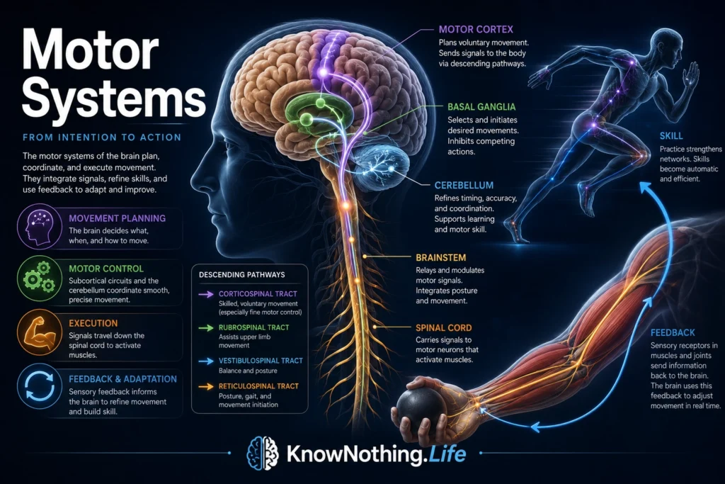
Motor systems are the neural networks that allow the body to move. They include the brain regions, spinal pathways, peripheral nerves, sensory feedback loops, and muscles that turn intention into action. Every voluntary movement, from reaching for a cup to speaking a sentence, depends on motor systems. So do many automatic and rhythmic actions, such as posture adjustment, balance correction, walking, swallowing, blinking, and breathing. The motor system is not one single pathway or one brain region. It is a coordinated set of cortical, subcortical, brainstem, spinal, and sensory circuits that work together to produce movement.
Movement may seem simple because it feels immediate, but it is one of the nervous system’s most complex achievements. A person lifting a hand must select the goal, plan the movement, activate the right muscles, inhibit competing muscles, adjust posture, predict sensory consequences, monitor feedback, correct errors, and stop the action at the right time. Motor systems therefore do far more than “send commands.” They organize the body in relation to the world. They translate perception, intention, motivation, and learned skill into controlled physical behavior.
Motor Cortex and Voluntary Movement
The motor cortex is a major cortical control system for voluntary movement. It lies in the frontal lobe, just anterior to the central sulcus, and includes the primary motor cortex, premotor cortex, and supplementary motor area. A StatPearls overview describes the motor cortex as a frontal-lobe region whose primary function is to send signals that direct the body’s movement. It also identifies the primary motor cortex, premotor cortex, and supplementary motor area as major components of motor cortical function.
The primary motor cortex, located mainly in the precentral gyrus, is strongly involved in generating voluntary motor output. The premotor cortex helps prepare movements in relation to external cues, while the supplementary motor area is especially important for internally generated actions, sequences, and coordinated movement plans. This division is not absolute, because real movement depends on overlapping networks. Still, it helps explain why the motor cortex is not just an “execution strip.” It participates in planning, selection, timing, and control. The brain must decide not only which muscle to move, but why, when, how strongly, and in what sequence.
The Motor Homunculus and Body Maps
One of the most famous ideas in motor neuroscience is the motor homunculus, a distorted body map showing how different body parts are represented along the motor cortex. Wilder Penfield and Edwin Boldrey helped popularize this image through cortical stimulation studies in neurosurgical patients. Their 1937 work remains historically important because it showed that movement-related cortical representation is organized, but uneven. Body parts requiring fine control, such as the hands, face, lips, and tongue, occupy proportionally large cortical regions. Modern reviews describe Penfield and Boldrey’s homunculus as a landmark in the mapping of human sensorimotor cortex.
The homunculus is useful, but it should not be treated as a perfectly literal map. Modern motor neuroscience shows that body representations are more distributed, overlapping, and network-based than the classic diagram suggests. Movements are not controlled one muscle at a time from isolated cortical points. Reaching, grasping, speaking, walking, and writing require coordinated patterns across multiple muscles and brain regions. The homunculus remains valuable because it captures an important truth: the brain represents the body according to functional importance, not physical size.
The Corticospinal Tract
The corticospinal tract is one of the most important descending motor pathways. It carries signals from the cerebral cortex through the brainstem and spinal cord to influence motor neurons that control muscles. StatPearls describes the corticospinal tract, also called the pyramidal tract, as the major neuronal pathway for voluntary motor function, connecting the cortex to the spinal cord and enabling movement, especially of the distal extremities.
A large portion of corticospinal fibers cross to the opposite side in the medulla, which helps explain why one hemisphere of the brain often controls movement on the opposite side of the body. Damage to this pathway can produce weakness, impaired fine motor control, spasticity, abnormal reflexes, or other upper motor neuron signs. The corticospinal tract is especially important for skilled voluntary movement of the hands and fingers. It allows the cortex to exert refined influence over spinal motor circuits, helping make tool use, writing, typing, surgery, drawing, and musical performance possible.
Basal Ganglia and Action Selection
The basal ganglia are deep brain structures involved in movement, habit, reward, motivation, and action selection. They do not simply start movement like an on-switch. Instead, they help determine which actions should be facilitated and which should be suppressed. Garrett Alexander, Mahlon DeLong, and Peter Strick proposed an influential model of parallel, functionally segregated circuits linking the basal ganglia and cortex, showing that basal ganglia loops participate not only in motor control but also in cognitive and limbic functions.
This action-selection role helps explain why basal ganglia disorders can produce very different motor symptoms. Parkinson’s disease can involve slowness, rigidity, tremor, and difficulty initiating movement. Huntington’s disease can involve involuntary movements and impaired control. Tourette syndrome, dystonia, and some compulsive behaviors also involve basal-ganglia-related circuits. The basal ganglia help the nervous system choose, initiate, scale, and sequence behavior. They are central to the difference between wanting to move and being able to release the right movement at the right moment.
Cerebellum and Movement Correction
The cerebellum is essential for coordination, timing, balance, motor learning, and error correction. It receives sensory information, motor plans, and feedback from many systems, then helps refine movement. A consensus paper on the cerebellum describes its roles in timing, sensory acquisition, and prediction of the sensory consequences of action.
The cerebellum does not usually cause paralysis when damaged. Instead, cerebellar injury tends to produce ataxia, tremor, poor coordination, inaccurate reaching, unstable posture, slurred speech, and difficulty with timing. This shows that movement requires more than muscle activation. It requires prediction and correction. The cerebellum helps compare intended movement with actual sensory feedback, allowing the nervous system to reduce error over time. When someone learns to ride a bike, throw a ball, play piano, or adjust to new glasses, cerebellar learning helps the body recalibrate movement.
Brainstem, Spinal Cord, and Reflex Control
Motor systems also depend heavily on the brainstem and spinal cord. The brainstem helps regulate posture, balance, eye movements, facial movements, swallowing, breathing, and many automatic motor functions. Descending brainstem pathways influence muscle tone and posture, while cranial nerve motor nuclei control muscles of the face, eyes, tongue, throat, and neck. The spinal cord contains motor neurons that directly activate skeletal muscles, as well as interneuron networks that coordinate reflexes and patterned movement.
Charles Sherrington’s 1906 work The Integrative Action of the Nervous System helped establish the nervous system as an integrated system of reflexes, inhibition, and coordinated action. Later reviews emphasize that Sherrington treated the synapse and reflex as central to understanding how elementary actions combine into complex behavior. Reflexes are not primitive mistakes beneath voluntary action. They are fast protective and regulatory systems. Withdrawal reflexes protect against harm, stretch reflexes help maintain posture, and spinal circuits contribute to rhythmic movement. Higher motor control builds on these lower-level systems rather than replacing them.
Central Pattern Generators and Rhythmic Movement
Some movements are rhythmic and patterned, such as walking, swimming, breathing, and chewing. These actions depend partly on central pattern generators, or CPGs, which are neural circuits capable of producing rhythmic motor output. A review on locomotor central pattern generators describes walking, flying, and swimming as largely controlled by spinal neuron networks known as CPGs.
CPGs do not make movement fully automatic in the sense of being disconnected from the brain. Descending control, sensory feedback, motivation, and environmental demands can modify their output. Walking across flat ground differs from walking upstairs, stepping over a puddle, or recovering from a stumble. The spinal cord can generate basic rhythmic structure, while the brain and sensory systems adapt that rhythm to real-world conditions. This layered organization makes movement efficient: the brain does not need to micromanage every muscle contraction, but it can guide, correct, and reshape movement as needed.
Sensory Feedback and Motor Learning
Motor systems depend on sensory feedback. Vision tells the brain where targets are. Proprioception tells it where limbs are. Touch tells it whether an object has been grasped. Vestibular input helps maintain balance. Pain warns that a movement may be harmful. Movement is therefore sensorimotor, not purely motor. The nervous system must constantly compare predicted outcomes with actual outcomes.
Motor learning occurs when repeated practice changes the nervous system. Basal ganglia circuits help reinforce useful actions and habits. The cerebellum helps reduce prediction errors and refine timing. Motor cortex and spinal circuits can reorganize with training. A review of basal ganglia and cerebellar loops notes that damage to basal ganglia or cerebellar components of motor circuits can produce motor symptoms, while related nonmotor loops contribute to higher-order deficits. This helps explain why learning movement is not merely building stronger muscles. It is also building better neural control.
Clinical Importance of Motor Systems
Motor-system disorders can arise from damage to the cortex, corticospinal tract, basal ganglia, cerebellum, brainstem, spinal cord, peripheral nerves, neuromuscular junction, or muscles. Stroke may damage motor cortex or descending pathways. Parkinson’s disease affects basal ganglia circuits. Cerebellar disorders impair coordination. Spinal cord injuries interrupt descending and ascending communication. Peripheral neuropathies weaken or distort signals to muscles. Motor neuron diseases damage the neurons that directly control muscle activity.
Clinical examination of movement can reveal where the nervous system is failing. Weakness, spasticity, tremor, rigidity, ataxia, fasciculations, abnormal reflexes, poor coordination, or gait disturbance all point to different levels of motor-system dysfunction. Motor rehabilitation depends on plasticity, practice, feedback, repetition, and motivation. Recovery is possible because motor systems are adaptive, but it is shaped by the location and severity of injury, the remaining pathways, and the quality of retraining.
Why Motor Systems Matter
Motor systems matter because movement is how the nervous system becomes visible in the world. Perception matters because it guides action. Emotion matters because it prepares action. Motivation matters because it selects action. Memory matters because it improves action. A brain that could sense and think but not move would be trapped inside itself. Motor systems allow intention to become behavior.
The deeper lesson is that movement is not separate from mind. Gesture, speech, posture, facial expression, tool use, writing, dance, sport, and touch all depend on motor control. Human intelligence is embodied because thinking evolved in organisms that had to move through space, reach for goals, avoid danger, and interact with others. To understand motor systems is to understand how the brain turns plans into motion, motion into skill, and skill into lived action.


