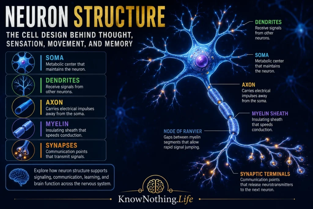
A neuron is a specialized cell of the nervous system designed to receive, process, and transmit information. Neurons are the basic signaling units of the brain, spinal cord, and peripheral nerves. They allow the body to sense the world, move muscles, form memories, regulate internal organs, feel pain, interpret emotion, and coordinate behavior. A typical neuron includes a cell body, dendrites, an axon, and synaptic terminals. The cell body contains the nucleus and supports the neuron’s metabolism; dendrites receive signals; the axon carries signals away; and synapses allow communication with other cells. A StatPearls overview summarizes this basic architecture by describing neurons as having a soma containing the nucleus, an axon, and a dendritic tree.
The neuron’s structure is important because form and function are inseparable in the nervous system. A neuron is not shaped randomly. Its branches, membranes, channels, axon length, myelin covering, and synaptic endings all support communication. Some neurons have short axons and participate in local circuits, while others have axons that extend long distances through the spinal cord or peripheral nerves. Some neurons have small dendritic trees, while others, such as Purkinje cells in the cerebellum, have enormous branching structures that receive vast amounts of input. The neuron is therefore both a cell and a communication device: a living biological unit built to integrate signals and influence other cells.
The Cell Body: The Neuron’s Metabolic Center
The cell body, also called the soma, is the central part of the neuron. It contains the nucleus, which houses the cell’s genetic material, and many organelles required for survival and function. These include mitochondria for energy production, ribosomes for protein synthesis, the endoplasmic reticulum for protein and lipid processing, and the Golgi apparatus for packaging and transporting cellular products. The soma keeps the neuron alive, maintains its internal chemistry, and produces many of the molecules needed for signaling, growth, repair, and synaptic communication. StatPearls describes the neuronal cell body as containing the nucleus and serving as a major site of metabolic activity.
The soma is also a place where incoming signals can influence whether the neuron will fire. Dendrites and synapses may bring many excitatory and inhibitory signals toward the cell body. These inputs are integrated across the neuron’s membrane, especially near the axon hillock, where the decision to generate an action potential is often made. The cell body is therefore not merely a maintenance station. It helps connect the neuron’s biological needs with its signaling role. A healthy soma allows the neuron to maintain ion gradients, synthesize proteins, transport materials, respond to injury, and participate in circuit activity.
Dendrites: The Neuron’s Receiving Branches
Dendrites are branching extensions that receive input from other neurons. Their tree-like shape allows a neuron to gather signals from many sources at once. Some dendrites receive only a modest number of synaptic contacts, while others receive thousands. This branching architecture is one reason neurons can participate in complex information processing. A neuron may receive signals related to sensation, memory, attention, emotion, or movement, then combine those signals into a response. NCBI’s Developmental Biology text describes dendrites as fine extensions used to pick up electrical impulses from other cells and notes that some neurons develop extensive dendritic trees.
Dendrites are not passive wires. They contain receptors, ion channels, biochemical signaling systems, and often small protrusions called dendritic spines. These spines are major sites of excitatory synapses in many neurons and can change shape with learning and experience. This makes dendrites central to neural plasticity. When a person learns a skill, remembers an event, or adapts to repeated experience, changes in synaptic strength and dendritic structure may help reshape the circuit. Dendrites therefore represent the neuron’s openness to the world: they are the branching surfaces where other cells influence what the neuron becomes likely to do.
The Axon Hillock and Action Potential
The axon hillock is the region where the soma narrows into the axon. It is especially important because it often acts as the trigger zone for action potentials. A neuron receives many inputs, some excitatory and some inhibitory. If the combined signal reaches threshold at the axon hillock or initial segment, voltage-gated ion channels open and an action potential begins. This action potential is an all-or-none electrical impulse that travels down the axon. A StatPearls review describes action potentials as electrical impulses generated by changes in sodium and potassium gradients across the neuronal membrane.
This process gives neurons a powerful form of decision-making at the cellular level. The neuron does not respond equally to every input. It integrates signals over time and space, then fires only if the conditions are strong enough. In this way, the axon hillock helps convert graded incoming activity into a clear outgoing signal. The nervous system depends on this transformation. Sensory perception, reflexes, voluntary movement, and thought all require neurons to decide when to remain silent and when to send a signal forward.
The Axon: The Neuron’s Transmission Line
The axon is the long projection that carries action potentials away from the cell body toward other neurons, muscles, or glands. Axons can vary enormously in length. Some are extremely short and communicate within a local brain circuit. Others extend from the spinal cord to distant muscles in the body. The axon’s job is to transmit information reliably over distance. It does this through a specialized membrane containing ion channels that allow electrical signals to propagate. The Queensland Brain Institute describes the axon as the long, thin structure in which action potentials are generated and transmitted toward neurotransmitter release.
Axons also contain internal transport systems that move proteins, vesicles, mitochondria, and other materials between the soma and axon terminal. This transport is essential because the axon may be far from the cell body. Without axonal transport, the neuron could not maintain synapses, repair damage, or support long-distance communication. The axon is therefore not just a cable. It is a living extension of the cell, constantly supplied and maintained so that signals can travel accurately through the nervous system.
Myelin and Nodes of Ranvier
Many axons are covered by myelin, a fatty insulating sheath that increases the speed and efficiency of signal transmission. In the central nervous system, myelin is produced by oligodendrocytes. In the peripheral nervous system, it is produced by Schwann cells. Myelin does not cover the axon continuously. It is interrupted by small gaps called nodes of Ranvier. At these nodes, voltage-gated sodium channels are highly concentrated, allowing the action potential to jump from node to node in a process called saltatory conduction. NCBI’s neuroscience text explains that myelin acts as an electrical insulator and greatly speeds action-potential conduction.
The nodes of Ranvier are small but essential. StatPearls describes them as short, specialized regions of axonal membrane not insulated by myelin, with high concentrations of voltage-gated sodium channels needed for action-potential generation. This arrangement allows the nervous system to transmit signals rapidly without requiring axons to become enormous. Myelin is one reason human movement, speech, reflexes, and perception can happen with such speed. When myelin is damaged, as in multiple sclerosis or other demyelinating diseases, signals may slow, weaken, or fail, showing how crucial neuron structure is to normal function.
Synaptic Terminals and Communication
At the end of the axon are synaptic terminals, also called axon terminals or terminal boutons. These structures allow the neuron to communicate with another cell. When an action potential reaches the terminal, calcium channels open, calcium enters the terminal, and synaptic vesicles can release neurotransmitters into the synaptic cleft. Those neurotransmitters bind to receptors on the next cell, influencing whether that cell becomes more or less likely to fire. StatPearls describes synapses as involving neurons with cell bodies, axons, and dendrites, with neurotransmitter release and receptor binding enabling communication.
Synapses are central to learning, memory, emotion, and behavior because they are not fixed switches. Their strength can change. Repeated activity can make some synapses more effective and others less effective. This is one of the foundations of neural plasticity. A memory is not stored in one synapse alone, but changes in synaptic networks help encode experience. A skill becomes smoother partly because repeated practice changes communication among neurons. Synaptic terminals therefore represent the output side of the neuron, but also one of the main places where experience reshapes the nervous system.
The Neuron Doctrine and the Discovery of Neuron Structure
The modern understanding of neuron structure emerged through one of the great scientific debates in biology. In the late nineteenth and early twentieth centuries, Camillo Golgi and Santiago Ramón y Cajal used staining techniques to reveal the structure of nerve cells. Golgi developed a staining method that made individual neurons visible in remarkable detail, while Cajal used such methods to argue that the nervous system was made of separate cells rather than one continuous web. Golgi and Cajal shared the 1906 Nobel Prize in Physiology or Medicine for their work on the structure of the nervous system.
This debate led to the neuron doctrine, the principle that neurons are individual structural and functional units of the nervous system. A historical review notes that Golgi and Cajal shared the Nobel Prize but held sharply different views, with the neuron doctrine emphasizing the structural, functional, and developmental individuality of nerve cells. Cajal’s drawings of neurons remain famous because they captured not only the beauty of neural structure, but also its logic: dendrites receive, axons transmit, and neurons communicate across specialized contact points. Modern neuroscience has expanded far beyond Cajal’s era, but his insight remains foundational.
Types of Neurons
Not all neurons have the same shape. Sensory neurons carry information from the body toward the central nervous system. Motor neurons carry commands from the central nervous system to muscles and glands. Interneurons connect neurons within local circuits and are especially abundant in the brain and spinal cord. Structurally, neurons may be classified as multipolar, bipolar, unipolar, or pseudounipolar depending on the arrangement of their processes. Multipolar neurons, common in the central nervous system, have one axon and many dendrites. Bipolar neurons, found in certain sensory systems, have one axon and one dendritic process. Pseudounipolar neurons often carry sensory information from the body.
These differences show how neuron structure fits neuron function. A sensory neuron must carry information from receptors toward the spinal cord or brain. A motor neuron must project toward a muscle and influence contraction. A cortical pyramidal neuron must integrate many inputs and send signals to other brain regions. A cerebellar Purkinje cell must receive enormous input through a vast dendritic tree and help regulate movement timing. The nervous system’s power comes partly from this diversity. There is no single “generic neuron” doing every job. There are many specialized neuron forms, each shaped by its role in the circuit.
Why Neuron Structure Matters
Neuron structure matters because it explains how the nervous system communicates. Dendrites receive signals, the soma supports and integrates them, the axon hillock helps initiate action potentials, the axon carries signals, myelin speeds conduction, nodes of Ranvier regenerate impulses, and synaptic terminals communicate with other cells. Each part of the neuron contributes to the larger process of neural signaling. Thought, movement, sensation, memory, and emotion all depend on these microscopic structures working together.
The neuron also shows that the brain is both biological and informational. It is made of living cells, but those cells are organized to transmit meaning-bearing signals. A neuron uses membranes, ions, proteins, neurotransmitters, organelles, and branching architecture to participate in perception, action, learning, and consciousness. To understand neuron structure is to understand the basic design principle of the nervous system: the mind depends on cells built to receive, transform, and pass information through living networks.