
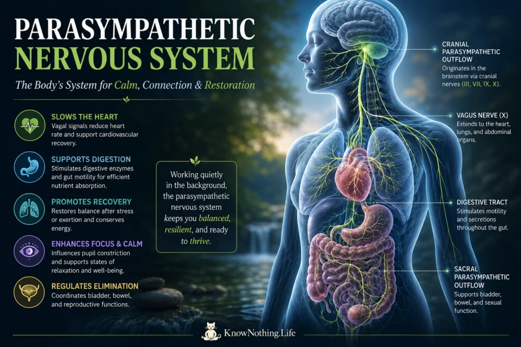
The parasympathetic nervous system is a major division of the autonomic nervous system, regulating organs and glands largely outside conscious awareness. It is commonly summarized as the body’s “rest-and-digest” system, but that phrase captures only part of its role. Parasympathetic pathways slow the heart under many conditions, support digestion and elimination, constrict the pupils, stimulate secretions, and help restore stability after exertion. Yet the system is not simply activated during relaxation. Human exercise studies show that vagal influence can persist at high workloads and becomes especially prominent during early recovery, demonstrating continuous adjustment rather than a simple switch between sympathetic and parasympathetic states.
Parasympathetic control is highly selective. The heart, airways, digestive tract, eyes, salivary glands, bladder, and reproductive organs do not receive one uniform command. Brainstem and spinal circuits instead generate patterned responses suited to breathing, eating, posture, sleep, emotion, and internal organ conditions. Experiments that selectively activated cholinergic neurons in the dorsal motor nucleus of the vagus produced pronounced slowing of the heart through peripheral acetylcholine receptors. Such findings show that defined parasympathetic circuits can control particular targets. The system is therefore better understood as a distributed regulatory network that conserves resources when appropriate while continually shaping organ function.
Anatomy and Cholinergic Signaling
Parasympathetic outflow is traditionally described as craniosacral. Preganglionic neurons in the brainstem send fibers through cranial nerves III, VII, IX, and X, while neurons in the sacral spinal cord supply pelvic organs. Tracing studies have identified brainstem neurons contributing parasympathetic fibers to the facial, glossopharyngeal, and vagus nerves, while separate experiments mapped sacral neurons projecting toward the bladder and lower bowel. The vagus has the broadest reach, traveling from the brainstem through the neck and thorax into the abdomen. Human anatomical work has also documented ascending parasympathetic nerves within the colon, showing that pelvic control is more extensive than simplified diagrams suggest.
Most parasympathetic pathways use a two-neuron chain. A relatively long preganglionic axon travels from the central nervous system to a ganglion near or inside the target organ, where it activates a short postganglionic neuron. Acetylcholine is the principal transmitter at both stages: nicotinic receptors mediate communication within autonomic ganglia, while muscarinic receptors usually produce the final organ response. Otto Loewi’s 1921 paper, Über humorale Übertragbarkeit der Herznervenwirkung, provided a landmark demonstration of chemical neurotransmission. Fluid transferred from a vagally stimulated frog heart slowed a second heart, revealing the action of the substance later identified as acetylcholine.
Cardiac and Respiratory Regulation
Parasympathetic control of the heart is carried mainly through the vagus nerve. Acetylcholine released near the sinoatrial node reduces pacemaker activity, while effects on the atrioventricular node slow conduction. At rest, vagal influence helps keep heart rate below the intrinsic rhythm generated by the heart’s pacemaker cells. When movement begins, rapid withdrawal of this inhibition allows heart rate to rise before slower sympathetic mechanisms become dominant. Pharmacological studies using atropine, which blocks muscarinic receptors, have shown that the earliest acceleration during light exercise is largely produced by vagal withdrawal rather than immediate sympathetic activation.
The return of heart rate toward baseline after exercise reflects renewed parasympathetic influence together with sympathetic withdrawal. In a controlled atropine study, parasympathetic effects were already substantial during the first minute of recovery and increased over the next several minutes. Christopher Cole and colleagues later reported in “Heart-Rate Recovery Immediately After Exercise as a Predictor of Mortality” that a delayed fall in heart rate after exercise, interpreted partly as reduced vagal reactivation, strongly predicted mortality after adjustment for exercise capacity and cardiac findings. Heart-rate recovery is not a pure measurement of one nerve pathway, but it demonstrates how restoration of parasympathetic control can provide clinically meaningful information about cardiovascular regulation.
Breathing is closely linked to this cardiac control. Heart rate normally rises during inhalation and falls during exhalation, producing respiratory sinus arrhythmia. Research involving patients with transplanted or denervated organs showed that feedback from the lungs and intact vagal pathways contribute to the rhythm, while arterial baroreceptors also shape it. Respiratory sinus arrhythmia and high-frequency heart-rate variability are therefore often used as estimates of cardiac parasympathetic modulation. However, pharmacological blockade studies show that these measures are affected by breathing rate, depth, posture, and individual physiology, so a larger heart-rate oscillation does not necessarily indicate greater parasympathetic activity throughout the body.
Digestion and the Gut–Brain Axis
The parasympathetic system participates in digestion through motor and sensory communication. Vagal motor pathways influence the stomach and upper digestive tract, coordinating smooth-muscle activity, secretion, and reflexes involved in processing a meal, while pelvic pathways contribute to the lower bowel. At the same time, many vagal fibers carry information from organs toward the brain rather than commands away from it. These sensory fibers detect stretch, nutrients, hormones, and chemical conditions, sending signals to the nucleus of the solitary tract. Parasympathetic function is therefore closely connected to interoception, the nervous system’s representation of the body’s internal state.
Experiments have clarified how this gut–brain communication influences eating. Selective removal of gut vagal and splanchnic sensory input altered the negative-feedback processes that normally promote satiation after nutrients enter the digestive tract, while leaving some forms of food reward intact. Other work found that gastrointestinal distension activated vagal afferents and brainstem circuits while reducing food intake and improving glucose tolerance in mice. Short-chain fatty acids produced during microbial fermentation have also been shown to suppress feeding partly through vagal sensory neurons. The vagus is thus not simply a digestive accelerator, but a bidirectional channel connecting meals, metabolism, behavior, and brain function.
Eyes, Glands, and Pelvic Organs
Cranial parasympathetic pathways regulate several visible functions. Fibers traveling with the oculomotor nerve activate the iris sphincter, constricting the pupil in bright light, and the ciliary muscle, which changes lens shape for near vision. Facial and glossopharyngeal pathways stimulate lacrimal and salivary glands. Human pharmacological experiments show that pupil changes during attention are not controlled solely by sympathetic dilation. Blocking the parasympathetic iris sphincter altered task-related responses, and later studies found that rapid pupil dilation can begin through reduced parasympathetic constrictor activity before slower sympathetic effects become prominent. Pupil size therefore reflects interaction between both autonomic divisions.
Sacral and pelvic circuits regulate urination, defecation, and aspects of sexual function. Parasympathetic activation contracts the bladder’s detrusor muscle and helps coordinate voiding, while pelvic pathways influence the distal colon and rectum. Viral-tracing and pharmacological experiments involving Barrington’s nucleus, a brainstem region associated with urination, demonstrated connections capable of coordinating colonic pressure through lumbosacral parasympathetic neurons. Human microscopic studies have also identified autonomic neurons within the bladder wall with parasympathetic characteristics. Normal elimination depends on cooperation among parasympathetic, sympathetic, sensory, and somatic motor pathways rather than the action of one isolated branch.
Inflammation, Dysfunction, and Neuromodulation
Parasympathetic pathways can influence immune activity through neural reflexes. In the landmark 2000 study “Vagus Nerve Stimulation Attenuates the Systemic Inflammatory Response to Endotoxin,” Linda Borovikova and colleagues found that electrical vagus stimulation reduced tumor necrosis factor production and protected animals from severe inflammatory responses. Later experiments showed that vagal sensory activation can recruit an anti-inflammatory pathway involving splanchnic sympathetic nerves, suggesting that neuroimmune regulation depends on connected circuits rather than a single direct route. This work helped establish bioelectronic medicine, which seeks to modify disease-related signaling by stimulating defined nerves.
Parasympathetic dysfunction can occur in diabetes, neurodegenerative disease, nerve injury, autoimmune disorders, and after surgery. Symptoms may include abnormal heart-rate responses, impaired digestion, dry mouth, urinary difficulties, pupillary abnormalities, or reduced exercise recovery. Clinical testing in diabetes found parasympathetic impairment more frequently than sympathetic impairment, with beat-to-beat heart-rate variation providing a sensitive indicator. Vagus nerve stimulation has also become an important neuromodulation strategy, and controlled studies in medication-resistant epilepsy have reported reductions in seizure frequency for some patients. Such therapies do not merely create generalized calm; they alter specific sensory and motor circuits linking the brain, organs, and immune system.


