
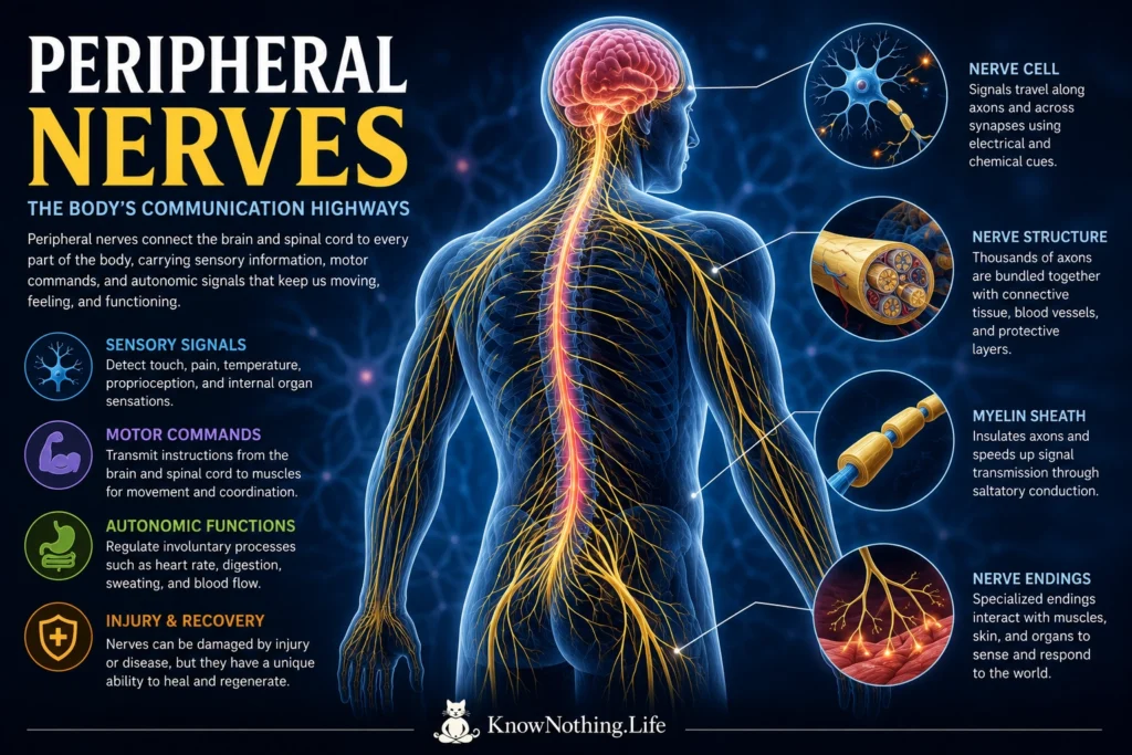
Peripheral nerves form the communication routes between the central nervous system and nearly every other part of the body. They carry sensory information from the skin, joints, muscles, and internal organs toward the brain and spinal cord, while motor fibers transmit commands back to skeletal muscles. Other peripheral fibers regulate involuntary processes involving blood vessels, sweat glands, digestion, and internal organs. A named nerve is therefore not a single, uniform wire. It may contain thousands of sensory, motor, and autonomic axons traveling together, each carrying a particular kind of information toward a specific destination.
This distributed network allows the nervous system to transform physical events into perception and action. Cutaneous receptors signal pressure, vibration, temperature, and potentially damaging stimulation. Muscle and joint receptors provide information about body position, movement, and force, while motor axons activate muscles. Human recordings from dorsal roots and cutaneous nerves have identified distinct fiber groups based on their diameter and conduction velocity. These findings demonstrate that peripheral nerves contain multiple signaling channels whose physical structure is closely related to their function.
The Internal Structure of Peripheral Nerves
A peripheral nerve is built from axons, glial cells, blood vessels, and several layers of connective tissue. Individual axons lie within delicate endoneurial tissue and are gathered into bundles called fascicles. Each fascicle is enclosed by the perineurium, while the entire nerve is surrounded by the tougher epineurium. These layers protect the axons, maintain the internal chemical environment, and allow nerves to tolerate a limited amount of movement and stretching. Sydney Sunderland’s influential 1951 paper, A Classification of Peripheral Nerve Injuries Producing Loss of Function, connected damage to these structural layers with the likelihood of neurological recovery.
Schwann cells are the principal supporting cells of peripheral nerves. Myelinating Schwann cells wrap repeatedly around larger axons to create a multilayered sheath, while nonmyelinating Schwann cells support groups of smaller axons without producing thick myelin. Myelin limits electrical current loss and allows impulses to be regenerated at specialized gaps called nodes of Ranvier. In their landmark 1949 study, Evidence for Saltatory Conduction in Peripheral Myelinated Nerve Fibres, Andrew Huxley and Robert Stämpfli showed that excitation advances between successive nodes. This arrangement greatly increases conduction speed while reducing the amount of membrane that must actively exchange ions.
How Peripheral Nerves Carry Electrical Signals
Peripheral axons transmit information through action potentials, brief electrical changes created by the movement of ions across the cell membrane. When an axon reaches threshold, voltage-sensitive sodium channels open and sodium ions enter, rapidly changing the membrane voltage. Potassium channels then contribute to repolarization and restoration of the resting state. Alan Hodgkin and Andrew Huxley established the mathematical foundations of this process in their 1952 paper, A Quantitative Description of Membrane Current and Its Application to Conduction and Excitation in Nerve. Although their experiments examined the squid giant axon, the principles became central to understanding neural excitability throughout the nervous system.
Signal speed depends heavily on axonal diameter, myelination, internodal distance, and temperature. Large, heavily myelinated axons generally conduct impulses faster than small or unmyelinated axons. Joseph Erlanger and Herbert Gasser’s early recordings showed that a mixed peripheral nerve produces several electrical components because its fibers conduct at different speeds. Subsequent anatomical and physiological studies confirmed a close relationship between fiber diameter and conduction velocity. Fast fibers carry motor commands, vibration, and proprioceptive signals, while smaller fibers contribute to temperature sensation, pain, itch, and autonomic regulation.
Sensation, Movement, and Reflexes
Sensory nerve endings are specialized to encode different aspects of the environment. Human microneurography experiments have identified several classes of fast-conducting mechanoreceptive fibers in the skin, each with characteristic receptive fields and response patterns. Some respond strongly when stimulation begins or changes, making them useful for detecting movement and vibration. Others continue firing during steady pressure and contribute to the perception of shape, edges, and sustained contact. Slowly conducting unmyelinated tactile fibers have also been recorded in human skin, showing that gentle touch is represented by more than one class of peripheral pathway.
Proprioceptive fibers provide information about the position and movement of the limbs. Muscle spindles respond to changes in muscle length, while tendon-related receptors help encode force. Direct recordings from human muscle-spindle afferents have shown that their firing changes during ankle, finger, and other voluntary movements. The central nervous system combines this feedback with descending motor commands to correct movements while they are occurring. Reflex pathways can also use peripheral sensory signals to produce rapid muscle responses through spinal circuits, helping preserve posture and protect tissues without waiting for deliberate conscious action.
Motor and Autonomic Fibers
Motor axons leave the spinal cord through ventral roots and travel through peripheral nerves before ending at neuromuscular junctions. There, acetylcholine released from the axon terminal activates receptors on the muscle membrane and initiates contraction. One motor neuron and all the muscle fibers it controls form a motor unit. Muscles used for precise movements generally contain many relatively small motor units, while muscles designed to generate greater force can contain larger units. Electrophysiological techniques that estimate motor-unit numbers are used to investigate nerve injury, aging, and disorders involving denervation.
Peripheral nerves also contain autonomic fibers serving smooth muscle, glands, blood vessels, the heart, and internal organs. These fibers help regulate sweating, circulation, digestion, pupil size, bladder activity, and other processes that usually occur without conscious direction. Damage to different fiber populations produces different clinical patterns. Large-fiber injury may cause weakness, loss of reflexes, poor balance, or reduced vibration and position sense. Small-fiber injury may produce burning pain, abnormal temperature perception, altered sweating, or cardiovascular and digestive symptoms, sometimes while standard nerve-conduction results remain normal.
Peripheral Nerve Injury and Wallerian Degeneration
Peripheral nerves can be damaged by compression, stretching, laceration, loss of blood supply, metabolic disease, inflammation, infection, and toxins. Hugh Seddon divided traumatic nerve injuries into neurapraxia, axonotmesis, and neurotmesis. Neurapraxia is a temporary conduction block without complete axonal disruption. Axonotmesis involves interruption of the axon with varying preservation of its surrounding pathways, while neurotmesis describes complete disruption of the nerve. Sunderland later expanded this system into multiple degrees based on which connective-tissue layers remained intact. These classifications remain useful because preserved internal pathways can guide axons toward their original targets.
When an axon is separated from its cell body, the distal portion undergoes Wallerian degeneration. The axon and its myelin break apart, Schwann cells change their behavior, and immune cells enter the damaged region to remove debris. This process is destructive, but it also prepares the distal nerve for regeneration. Schwann cells form aligned structures traditionally called bands of Büngner, which provide pathways for growing axons. Experimental studies have shown that migrating Schwann cells help organize cellular bridges across injury gaps and direct regenerating axons toward the distal stump.
Regeneration and the Limits of Recovery
Peripheral axons can regenerate more effectively than most axons in the brain and spinal cord, but recovery is neither automatic nor complete. New axonal sprouts must cross the damaged area, enter appropriate pathways, grow over potentially long distances, and reconnect with muscles or sensory receptors. Seddon, Peter Medawar, and H. Smith’s 1943 study, Rate of Regeneration of Peripheral Nerves in Man, helped establish clinical observations about human nerve regrowth. The commonly cited estimate of approximately one millimeter per day is only a rough guide because age, injury severity, distance, scarring, blood supply, and repair timing can all affect progress.
Regenerating axons may also enter the wrong fascicles or reconnect with inappropriate targets. A motor axon can reach the wrong muscle, while sensory fibers may establish altered receptive fields. These errors can leave persistent weakness, numbness, impaired coordination, or neuropathic pain even after anatomical continuity has been restored. Human microneurography studies have found abnormal receptive fields and response properties in regenerating sensory fibers. A controlled clinical study of digital-nerve repair also found that brief postoperative electrical stimulation improved measures of sensory recovery, suggesting that neural activity can influence regeneration.
Peripheral Neuropathy and Clinical Diagnosis
Peripheral neuropathy may affect one nerve, several separate nerves, or many nerves in a symmetrical pattern. Causes include diabetes, hereditary disorders, autoimmune disease, infection, nutritional deficiency, kidney disease, medications, toxins, and mechanical entrapment. The longest axons are often affected first, producing symptoms that begin in the feet and gradually move upward. Longitudinal diabetes studies have documented progressive abnormalities in sensory and motor nerve conduction and have associated sustained hyperglycemia with declining peripheral nerve function. Sensory fibers may deteriorate before major motor abnormalities become clinically apparent.
Nerve-conduction studies measure the speed and amplitude of responses in large myelinated fibers, while electromyography detects changes in muscles caused by motor axon loss and reinnervation. These methods can help distinguish axonal damage from demyelination and identify focal entrapment, but they may miss disorders confined to small fibers. Skin biopsy can quantify intraepidermal nerve endings, and large multicenter studies have established reference values that account for age and other variables. High-resolution ultrasound can reveal nerve enlargement, compression, discontinuity, or scarring, complementing physiological testing with structural information. Together, these methods show that peripheral nerves are living, adaptable tissues rather than passive electrical cables.


