
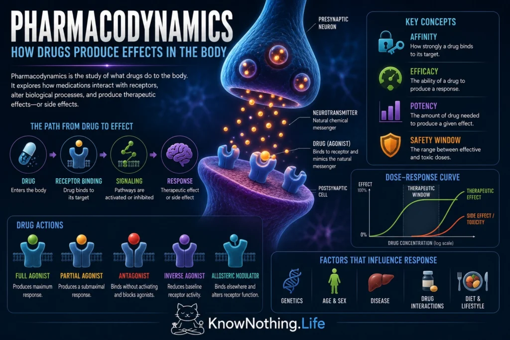
Pharmacodynamics is the study of how drugs affect cells, tissues, organs, and behavior. It examines the relationship between drug concentration and biological response, including the molecular targets a drug binds, the signaling pathways it changes, the strength of the resulting effect, and the adverse effects that may appear as exposure increases. Pharmacokinetics asks how the body absorbs, distributes, metabolizes, and eliminates a drug; pharmacodynamics asks what happens when that drug reaches its target.
The same concentration can produce different outcomes in different tissues because cells do not all express the same receptors or signaling machinery. A drug may reduce pain in one neural circuit, slow breathing in another, and alter digestion elsewhere. Pharmacodynamic models therefore connect receptor binding with the response of a complete biological system. The operational model introduced by James Black and Paul Leff in 1983 formalized this relationship by accounting for both a drug’s properties and the efficiency with which a particular tissue converts receptor activation into an observable effect.
Receptors and Signal Transduction
Many drugs produce their effects by binding to receptors, proteins that recognize particular chemical structures and initiate or prevent cellular responses. Receptors may be located on the cell membrane, within the cytoplasm, or inside the nucleus. Some open ion channels within milliseconds, while others activate G proteins, enzymes, second messengers, or changes in gene expression. Drugs can also act on transporters, enzymes, structural proteins, and voltage-sensitive channels, so receptor binding is central to pharmacodynamics but does not explain every drug action.
Early receptor theories assumed that biological response was directly proportional to the number of occupied receptors. This simple idea could not explain why two drugs occupying the same receptor population might produce different maximum effects. In Affinity and Intrinsic Activity in the Theory of Competitive Inhibition, E. J. Ariëns separated the ability to bind a receptor from the ability to activate it. R. P. Stephenson expanded this distinction in his 1956 paper A Modification of Receptor Theory, arguing that receptor occupancy and tissue response need not be identical. These concepts became the foundation for modern distinctions among affinity, efficacy, and potency.
Affinity, Efficacy, and Potency
Affinity describes how strongly a drug binds to its target. A high-affinity compound can occupy receptors at relatively low concentrations, but strong binding does not guarantee a strong biological effect. Efficacy describes the ability of the receptor-bound drug to produce a response. A drug may bind tightly yet activate the receptor weakly, while another may have lower affinity but generate a greater maximum effect once enough of it reaches the target. These properties help explain why binding measurements alone cannot fully predict clinical performance.
Potency refers to how much drug is required to produce a specified effect and is commonly described using an EC50, the concentration producing half of the maximum observed response. Potency depends on affinity, efficacy, receptor density, and the efficiency of downstream signaling. A more potent drug is not necessarily more effective or safer; it may simply achieve a given response at a lower concentration. Black and Leff’s operational model showed why the apparent potency and efficacy of an agonist can change when it is tested in tissues with different receptor numbers or signal-amplification systems.
Receptor Reserve and Maximum Response
Some tissues contain more receptors than are required to produce a maximum response. These surplus binding sites are often called spare receptors or receptor reserve. In such a system, a full response may occur while only a fraction of the available receptors are occupied. As a result, the concentration required to produce half of the maximum response can be substantially lower than the concentration needed to occupy half of the receptors. Receptor reserve allows tissues to remain responsive even when receptor numbers decline or when an agonist has limited efficacy.
Experiments using irreversible antagonists have helped demonstrate receptor reserve. By permanently inactivating portions of a receptor population, researchers can observe whether the maximum response declines immediately or remains intact until a substantial number of receptors have been removed. Studies of vascular alpha-adrenoceptors found that some receptor populations retained strong responses despite progressive receptor inactivation, whereas others showed little or no reserve. Receptor reserve is therefore a property of the drug–receptor–tissue system rather than a permanent feature of the receptor alone.
Full, Partial, and Inverse Agonists
An agonist binds to a receptor and promotes an active signaling state. A full agonist can produce the maximum response available in a particular experimental system, while a partial agonist produces a lower maximum even when it occupies many or all available receptors. Partial agonists can behave differently depending on the surrounding level of receptor activity. In a system with little natural stimulation, they may increase signaling; in the presence of a strong full agonist, they may reduce the overall response by competing for the same receptors.
Buprenorphine provides a clinically important example. It acts as a partial agonist at the mu-opioid receptor and displays a plateau in several opioid effects as doses increase. In controlled human studies, subjective opioid effects and respiratory depression eventually increased less steeply than they did with full agonists such as methadone or fentanyl. This ceiling does not make buprenorphine incapable of causing respiratory harm, especially when combined with other depressants, but it illustrates how limited intrinsic efficacy can shape a drug’s pharmacodynamic profile.
Some receptors also display activity even when no agonist is bound. An inverse agonist binds to such a receptor and reduces its activity below the unoccupied baseline. A neutral antagonist, by contrast, blocks other ligands without substantially changing baseline signaling. Experiments involving beta-adrenergic receptors showed that several compounds traditionally classified as antagonists could reduce spontaneous receptor activity, demonstrating inverse agonism and expanding drug classification beyond a simple agonist-versus-antagonist model.
Competitive and Noncompetitive Antagonism
A competitive antagonist binds reversibly to the same receptor site as an agonist but does not activate it. Increasing the agonist concentration can overcome this blockade because the two molecules compete for the same binding site. On a concentration-response graph, a simple competitive antagonist shifts the agonist curve to the right without necessarily reducing the maximum response. The agonist becomes less potent in the antagonist’s presence, but its efficacy can remain intact if a sufficiently high concentration is reached.
Orompicha Arunlakshana and Heinz Schild established a rigorous method for studying this process in their 1959 paper Some Quantitative Uses of Drug Antagonists. Schild analysis uses changes in the agonist dose ratio to estimate antagonist affinity and determine whether observed blockade is consistent with simple competitive action. Their approach became one of pharmacology’s most important methods for identifying and comparing receptors before molecular cloning made receptor proteins directly accessible.
Noncompetitive antagonists reduce signaling in ways that cannot be fully overcome by adding more agonist. They may bind irreversibly to the agonist site, block an ion channel, or attach to an allosteric site that stabilizes a less active receptor state. These mechanisms often reduce the maximum attainable response, although receptor reserve can initially conceal that reduction. The final concentration-response curve therefore reflects both the antagonist’s molecular action and the capacity of the tissue to amplify signals from the receptors that remain functional.
Dose–Response Relationships and Therapeutic Windows
A graded dose-response curve shows how the magnitude of an effect changes as drug concentration increases. At low concentrations, little response may be measurable. The effect then rises through a responsive range before approaching a maximum, commonly called Emax. Different drug effects can have different curves. A dose that produces a desired therapeutic response may simultaneously begin producing sedation, low blood pressure, impaired coordination, or another adverse effect through the same or a different target.
Human receptor-imaging studies show how this principle operates clinically. Lars Farde and colleagues used positron emission tomography to measure dopamine D2 receptor occupancy in people receiving antipsychotic medications. Classical antipsychotics commonly occupied a large proportion of striatal D2 receptors, and patients with movement-related adverse effects tended to have higher occupancy. A later double-blind study connected receptor occupancy with both antipsychotic response and extrapyramidal symptoms. These experiments showed that therapeutic and adverse effects may occupy overlapping but distinguishable portions of a pharmacodynamic response curve.
Time Delays and Indirect Drug Effects
Drug concentration and effect do not always rise and fall together. A drug may reach the bloodstream quickly but require time to enter its target tissue. Alternatively, target binding may occur immediately while the observable response develops slowly because proteins must be synthesized, physiological substances must be depleted, or downstream systems must reorganize. The effect may therefore continue increasing after the plasma concentration has begun to decline, producing a delay between measured exposure and response.
Warfarin illustrates an indirect pharmacodynamic effect. It inhibits vitamin K epoxide reductase, but anticoagulation depends on the gradual reduction of active vitamin K-dependent clotting factors already present in circulation. The concentration of warfarin and the measured international normalized ratio therefore follow different time courses. Population PK–PD research has modeled this delay and found that VKORC1 genotype influences pharmacodynamic sensitivity, while CYP2C9 and age affect the handling of the more potent warfarin enantiomer.
Tolerance, Desensitization, and Adaptation
Pharmacodynamic responses can change during repeated treatment even when drug concentrations remain similar. Receptors may become phosphorylated, uncouple from signaling proteins, move inside the cell, decline in number, or activate compensatory pathways. These adaptations can produce tolerance, meaning that the same concentration generates a smaller response than it did previously. Other systems may become sensitized, producing a larger response after repeated or intermittent exposure.
Laura Bohn and colleagues demonstrated one mechanism in mice lacking beta-arrestin-2, a protein involved in mu-opioid receptor regulation. Chronic morphine did not produce ordinary receptor desensitization or analgesic tolerance in these animals, although physical dependence still developed. The findings showed that tolerance and dependence are not identical processes and can arise through partly different cellular adaptations. Later studies have revealed additional pathways, emphasizing that pharmacodynamic tolerance depends on the drug, receptor, tissue, exposure pattern, and effect being measured.
Individual Differences in Drug Response
People receiving the same drug concentration may experience different effects because pharmacodynamics varies among individuals. Receptor expression, genetic variation, age, disease, hormonal state, previous treatment, and interacting medications can all change target sensitivity. Some differences occur downstream from drug concentration, meaning that even perfect pharmacokinetic dosing cannot guarantee an identical biological response.
Warfarin again provides a clear example. Variants in VKORC1 alter sensitivity at the drug’s molecular target, while CYP2C9 variants influence metabolism. Large studies combining genetic and clinical data have shown that these factors explain a meaningful portion of the variation in stable therapeutic dose. Rare VKORC1 variants can also produce pronounced pharmacodynamic resistance, requiring unusually high exposure to achieve anticoagulation. These findings demonstrate why effective dosing must account for both how much drug reaches the body and how strongly the body responds.
Why Pharmacodynamics Matters
Pharmacodynamics explains why increasing a dose may strengthen one effect, reveal another, or eventually add toxicity without providing further benefit. It distinguishes binding from activation, potency from efficacy, and receptor occupancy from whole-body response. It also shows why partial agonists, inverse agonists, antagonists, and allosteric modulators can produce very different outcomes even when they act within the same receptor system.
The field connects molecular pharmacology with clinical treatment. Receptor theory helps researchers design drugs, dose-response studies identify useful concentration ranges, imaging confirms target engagement, and pharmacodynamic monitoring reveals whether a patient is responding safely. The central question is not simply whether a drug reaches its target, but how that target and the surrounding biological system transform chemical binding into benefit, adaptation, or harm.


