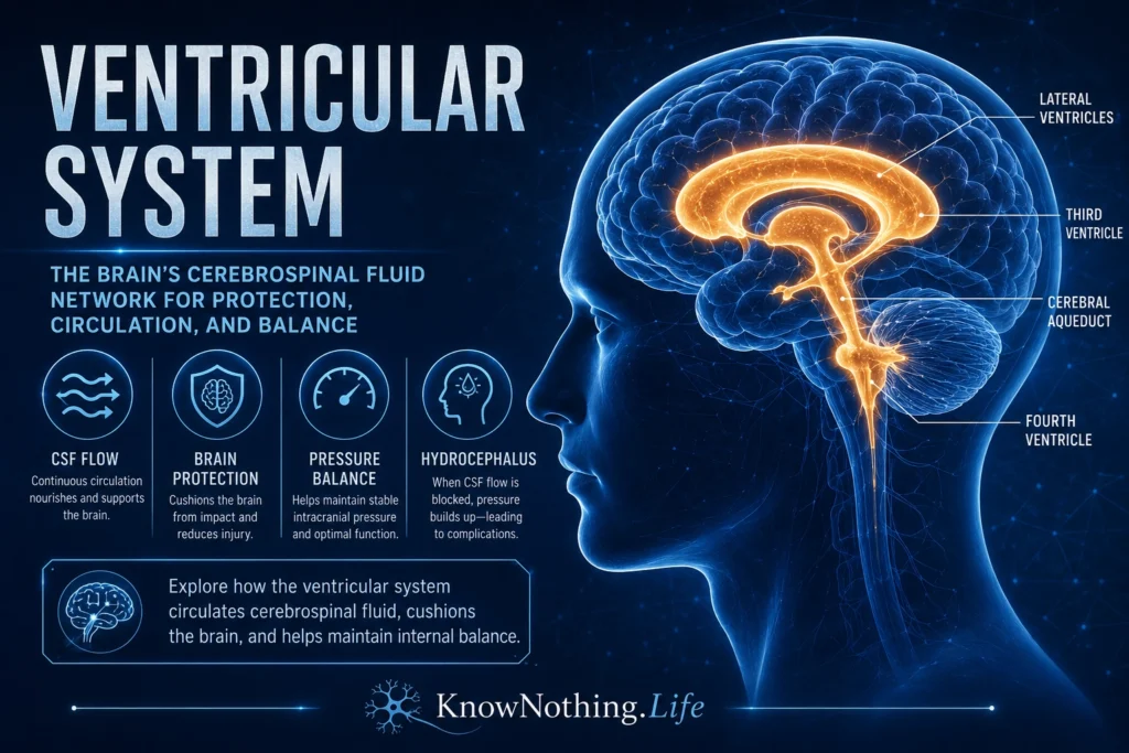
The ventricular system is a connected network of fluid-filled cavities deep inside the brain. These cavities, called ventricles, contain cerebrospinal fluid, or CSF, a clear fluid that cushions the brain and spinal cord, helps maintain chemical stability, transports nutrients and signaling molecules, and contributes to waste removal. The ventricular system includes two lateral ventricles, the third ventricle, the cerebral aqueduct, and the fourth ventricle. Together, they form a central fluid pathway that connects with the subarachnoid space surrounding the brain and spinal cord. StatPearls describes the ventricular system as an interconnected series of CSF-filled cavities that cushion the brain.
Although the ventricular system is often overlooked in popular descriptions of the brain, it is essential to normal nervous-system function. The brain is not a solid organ packed tightly into the skull. It is suspended, protected, and chemically supported within a fluid environment. The ventricles are part of that environment. They do not “think,” move muscles, or process language in the same way cortical and subcortical structures do, but they support the physical and biochemical conditions that make neural function possible. To understand the ventricular system is to understand that the brain depends not only on neurons, but also on circulation, pressure balance, fluid movement, and protective architecture.
The Four Ventricles of the Brain
The largest ventricles are the two lateral ventricles, one in each cerebral hemisphere. Each lateral ventricle has a curved shape and extends through parts of the frontal, parietal, occipital, and temporal regions. The lateral ventricles connect to the third ventricle through small openings called the interventricular foramina, also known as the foramina of Monro. The third ventricle is a narrow midline cavity located in the diencephalon, between the two sides of the thalamus and near the hypothalamus. From there, CSF passes through the cerebral aqueduct of Sylvius, a thin channel in the midbrain that connects the third ventricle to the fourth ventricle. The fourth ventricle lies between the brainstem and cerebellum and narrows downward toward the central canal of the spinal cord.
This anatomy matters because the ventricular system is both connected and vulnerable. A blockage at a narrow point, especially the cerebral aqueduct, can disrupt CSF movement and enlarge the ventricles upstream. The shape of the ventricles also helps clinicians interpret brain imaging. Enlarged ventricles may suggest hydrocephalus, brain-volume loss, obstruction, altered CSF absorption, or other neurological conditions. The ventricles are therefore not simply empty spaces. They are anatomical landmarks, fluid pathways, and clinical indicators of brain health.
Cerebrospinal Fluid and the Choroid Plexus
Cerebrospinal fluid is produced largely by the choroid plexus, a specialized tissue found in the lateral, third, and fourth ventricles. The choroid plexus contains vascular tissue and epithelial cells that help produce and regulate CSF. StatPearls notes that the majority of CSF is produced in the cerebral ventricles by the choroid plexus and ependymal cells. The choroid plexus is not merely a faucet pouring water into the brain. It helps control the composition of CSF, forming part of the blood-CSF barrier and influencing the chemical environment surrounding the central nervous system.
The amount of CSF in circulation is small compared with the total fluid volume produced across a day. Adult humans continuously produce and absorb CSF, with common estimates around several hundred milliliters per day. A StatPearls review of the choroid plexus notes that it may secrete up to about 500 ml of CSF per day in the adult human brain. This constant turnover shows that CSF is dynamic, not stagnant. It is made, circulated, exchanged, and reabsorbed, helping maintain the brain’s internal environment.
The Flow of Cerebrospinal Fluid
The traditional pathway of CSF flow begins in the lateral ventricles. From there, CSF travels through the interventricular foramina into the third ventricle, then through the cerebral aqueduct into the fourth ventricle. It exits the fourth ventricle through the median aperture of Magendie and the two lateral apertures of Luschka, entering the subarachnoid space around the brain and spinal cord. CSF also communicates with the central canal of the spinal cord. StatPearls summarizes this route from the lateral ventricles to the third ventricle, fourth ventricle, and subarachnoid space through the apertures of Magendie and Luschka.
This traditional model remains useful, but modern CSF research has become more complex. Some researchers have argued that the older view of CSF as simply produced by the choroid plexus, flowing unidirectionally through the ventricles, and being absorbed mainly by arachnoid granulations is incomplete. Miyajima and colleagues noted that the traditional CSF hydrodynamics hypothesis has been reconsidered in light of clinical and experimental observations. Newer models emphasize pulsatile flow, exchange with interstitial fluid, vascular influences, lymphatic drainage pathways, and local production or absorption dynamics. The important point is that CSF circulation is not just plumbing. It is a living fluid system shaped by pressure, pulsation, barriers, tissue exchange, and brain physiology.
What the Ventricular System Does
The ventricular system helps protect the brain physically. CSF provides buoyancy, reducing the effective weight of the brain and helping cushion it against sudden movement. Without fluid support, the brain’s soft tissue would be more vulnerable to compression and trauma. CSF also helps distribute forces within the skull, protecting delicate neural structures from some mechanical stress. This does not make the brain invulnerable, but it shows why fluid support is essential. The central nervous system is soft, metabolically demanding, and highly sensitive to pressure changes.
CSF also supports chemical stability. It helps maintain the extracellular environment in which neurons and glial cells function. The nervous system depends on tightly regulated concentrations of ions, nutrients, signaling molecules, and waste products. Reviews of the cranial meninges summarize major CSF functions as cushioning the brain and spinal cord, supplying nutrients, and removing waste. CSF also carries cytokines, growth factors, and extracellular vesicles, and research on cilia-driven flow in the ventricles notes that ventricular CSF contains biologically active molecules that move along ventricular walls with help from motile cilia. The ventricular system therefore contributes not only to protection, but also to communication and maintenance.
Ependymal Cells, Cilia, and Ventricular Walls
The ventricles are lined by ependymal cells, a specialized type of glial-related cell that forms a thin epithelial-like lining along the ventricular surface. These cells help separate brain tissue from ventricular CSF and contribute to the movement and regulation of fluid. Many ependymal cells possess cilia, tiny hair-like structures that beat rhythmically and help move CSF along ventricular surfaces. This ciliary motion is especially important during development and may help distribute molecular signals in the ventricular system.
The ventricular lining is not a passive wall. It is part of the brain’s fluid-interface system. During development, the ventricular zone is also a major region for neural progenitor cells, making the ventricular system important in brain formation as well as adult physiology. Korzh’s review on ventricular system development describes the brain ventricular system as consisting of ventricles and connecting channels filled with CSF, emphasizing its developmental significance. This means the ventricles are not simply spaces left over after the brain forms. They are part of the architecture around which the brain develops.
Hydrocephalus and Ventricular Enlargement
The most familiar clinical condition involving the ventricular system is hydrocephalus. Hydrocephalus occurs when CSF accumulates in the ventricles, often causing ventricular enlargement and increased pressure or impaired brain function. StatPearls defines hydrocephalus as symptomatic accumulation of CSF inside the cerebral ventricles. It can result from obstruction of CSF flow, impaired absorption, overproduction in rare cases, or altered CSF dynamics. Causes may include congenital malformations, hemorrhage, infection, tumors, trauma, or age-related changes.
Hydrocephalus can affect infants, children, and adults differently. In infants, the skull bones have not fully fused, so increased ventricular pressure may enlarge the head. In older children and adults, symptoms may include headache, nausea, vomiting, vision problems, balance difficulty, cognitive changes, urinary symptoms, or altered consciousness depending on severity and cause. Normal pressure hydrocephalus, typically seen in older adults, is associated with gait disturbance, urinary incontinence, and cognitive decline, though diagnosis and treatment require careful medical evaluation. Hydrocephalus shows why ventricular pressure and CSF movement matter: when the fluid system fails, brain function can be seriously affected.
Imaging and Clinical Importance
The ventricular system is highly visible on brain imaging, including CT and MRI scans. Clinicians use ventricular size, shape, symmetry, and surrounding tissue patterns to evaluate many conditions. Enlarged ventricles may reflect hydrocephalus, brain atrophy, obstruction, or developmental variation. A shift in ventricular position may suggest mass effect from bleeding, swelling, tumor, or other lesions. Blood inside the ventricles may indicate intraventricular hemorrhage. The ventricles also serve as landmarks for neurosurgical planning.
Treatments involving the ventricular system often focus on restoring CSF flow or reducing harmful accumulation. Hydrocephalus may be treated with a shunt system that diverts CSF to another body cavity, or with procedures such as endoscopic third ventriculostomy in selected cases. These interventions show that the ventricular system is not just an anatomical curiosity. It is a practical route for diagnosis, monitoring, and treatment. Neurosurgeons, neurologists, radiologists, pediatricians, and emergency physicians all rely on knowledge of ventricular anatomy.
Why the Ventricular System Matters
The ventricular system matters because it reveals the brain as a fluid-supported organ. Neurons may produce thought, memory, emotion, and movement, but they depend on a carefully regulated environment. CSF cushions the brain, supports chemical balance, transports molecules, contributes to waste clearance, and helps maintain pressure relationships inside the skull. The ventricles are central channels in that system. They remind us that brain function depends not only on electrical signals, but also on fluid circulation and physical protection.
The ventricular system also matters because small disruptions can have large consequences. A blocked aqueduct, abnormal CSF absorption, bleeding into the ventricles, or excessive ventricular enlargement can affect consciousness, movement, cognition, vision, and survival. To understand the ventricular system is to understand one of the brain’s essential support networks: a hidden fluid architecture that protects, nourishes, circulates, and stabilizes the nervous system from within.