
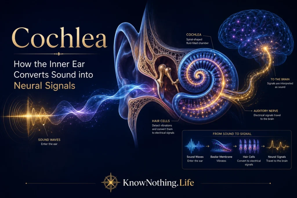
The cochlea is the spiral-shaped sensory organ of hearing located within the inner ear. Its name comes from the Latin word for snail, reflecting its coiled appearance. Although small enough to fit inside the temporal bone, the cochlea performs an extraordinary sequence of mechanical, electrical, and neural operations. It receives vibrations transmitted through the outer and middle ear, separates complex sounds into their frequency components, converts mechanical movement into electrical activity, and passes encoded information to the auditory nerve. Every spoken sentence, musical note, and environmental sound begins its neural journey through this compact biological system.
The cochlea is more than a microphone that responds whenever sound enters the ear. Its physical structure actively shapes auditory information before the brain receives it. Different regions respond preferentially to different frequencies, sensory cells adjust the sensitivity and sharpness of the response, and specialized synapses preserve the timing and intensity of rapidly changing sounds. The cochlea therefore performs the first major stage of auditory analysis. Damage to any of its interacting components—including hair cells, supporting cells, neural connections, blood supply, or fluid-regulating tissues—can change hearing even when other parts remain intact.
Cochlear Anatomy and the Organ of Corti
The cochlea contains three fluid-filled compartments known as the scala vestibuli, scala media, and scala tympani. The scala vestibuli and scala tympani contain perilymph, while the scala media contains potassium-rich endolymph. The basilar membrane separates the scala media from the scala tympani, and the organ of Corti rests on its upper surface. The organ of Corti contains the sensory hair cells of hearing together with supporting cells and specialized membranes that control how sound-induced vibrations reach the sensory receptors. The tectorial membrane lies above the hair cells and participates in the relative movement that bends their stereocilia.
The human organ of Corti contains one row of inner hair cells and approximately three rows of outer hair cells. These two receptor populations have very different functions. Inner hair cells provide most of the sensory information transmitted to the brain, while outer hair cells mechanically modify cochlear motion. Supporting structures keep these cells aligned and help maintain the precise geometry required for sound transduction. The spiral ganglion contains the cell bodies of auditory nerve neurons, whose peripheral fibers contact hair cells and whose central fibers form the cochlear portion of the eighth cranial nerve.
Traveling Waves and Cochlear Tonotopy
When the stapes bone moves against the oval window, it creates pressure changes within the cochlear fluids. These changes produce a traveling wave that moves along the cochlear partition. Georg von Békésy’s landmark investigations of inner-ear mechanics showed that different sound frequencies produce their largest mechanical effects at different places along the basilar membrane. High-frequency sounds reach maximum displacement near the stiff, narrow base of the cochlea, while lower-frequency sounds travel farther toward its wider and more flexible apex. Békésy presented these findings in works including Experiments in Hearing, establishing the physical basis of cochlear frequency analysis.
This orderly relationship between sound frequency and cochlear position is called tonotopy. It creates a frequency map that remains partly preserved throughout the auditory nerve, brainstem, thalamus, and auditory cortex. A complex sound containing many frequencies produces several overlapping patterns of cochlear activity rather than one uniform response. Recent measurements confirm that tonotopic mechanics extend through both basal and apical regions, although the low-frequency apex remains more difficult to study experimentally. The cochlea thus behaves like a biological filter bank, separating the acoustic spectrum into neural channels before central auditory processing begins.
Hair Cells and Mechanoelectrical Transduction
Hair cells receive their name from bundles of microscopic stereocilia projecting from their upper surfaces. Sound-induced movement within the organ of Corti bends these bundles, placing tension on fine links connecting neighboring stereocilia. This mechanical force opens ion channels near the stereociliary tips. Potassium and other ions flow into the hair cell from the endolymph, altering its membrane potential. Movement in the opposite direction reduces channel opening and produces the reverse electrical change. This conversion of mechanical force into an electrical receptor potential is known as mechanoelectrical transduction.
Research has identified TMC1 and TMC2 as essential components of the hair-cell transduction apparatus. Experiments by Beurg and colleagues showed that these proteins contribute to the properties of the mechanotransduction channel, while later work provided evidence that TMC1 forms part of its ion-conducting pore. Mutations affecting TMC1 can cause hereditary deafness because the hair cell may remain physically present but lose its ability to translate stereociliary movement into a normal electrical signal. These findings connect the molecular structure of hair cells with the fundamental act of hearing.
Outer Hair Cells and the Cochlear Amplifier
Outer hair cells act as mechanical effectors as well as sensory receptors. When their membrane voltage changes, they rapidly shorten or lengthen through a process called electromotility. This movement feeds energy back into the cochlear partition, increasing the vibration caused by quiet sounds and sharpening frequency selectivity. The resulting cochlear amplifier allows mammals to detect faint signals while distinguishing between frequencies that differ only slightly. It also produces compression, enabling the auditory system to respond across an enormous intensity range without treating every increase in sound pressure as an equally large increase in perceived loudness.
The motor protein prestin is central to outer-hair-cell electromotility. In 2000, Jing Zheng and colleagues identified prestin as the molecular motor responsible for rapid changes in outer-hair-cell length. A later experiment led by Joseph Liberman showed that mice lacking functional prestin lost outer-hair-cell electromotility and experienced a reduction of approximately 40 to 60 decibels in cochlear sensitivity, even though mechanotransduction within the cells remained present. These results demonstrated that outer hair cells do not simply detect sound; they actively enhance the mechanical conditions under which inner hair cells operate.
Inner Hair Cells, Ribbon Synapses, and the Auditory Nerve
Inner hair cells are the principal sensory output cells of the cochlea. When sound depolarizes an inner hair cell, voltage-gated calcium channels open near its basal surface. Calcium entry triggers the release of glutamate onto the endings of spiral ganglion neurons. The resulting activity travels through auditory nerve fibers toward the cochlear nuclei of the brainstem. Each inner hair cell contacts multiple nerve fibers, which differ in spontaneous activity, threshold, and intensity range. Their combined responses allow the auditory nerve to represent sounds extending from near the threshold of hearing to much higher levels.
Communication between an inner hair cell and an auditory nerve fiber occurs at a specialized ribbon synapse. A dense protein structure holds synaptic vesicles close to release sites, supporting fast and sustained neurotransmission. This design is essential because auditory signals contain timing differences measured in fractions of a millisecond. Tobias Moser and colleagues showed that calcium-dependent release at these synapses can preserve exceptional temporal precision, while experiments involving loss of the ribbon protein RIBEYE found impaired high-rate and precisely timed sound encoding. The ribbon synapse therefore converts the hair cell’s graded receptor potential into the precisely timed spikes used by the nervous system.
The Stria Vascularis and Cochlear Energy
Sensory transduction depends on the unusual electrical and chemical environment of the scala media. The stria vascularis, a highly vascularized tissue along the outer wall of the cochlear duct, maintains the potassium-rich endolymph and generates the positive endocochlear potential. This electrical gradient supplies much of the driving force that pushes ions into hair cells when their mechanotransduction channels open. The stria vascularis can therefore be understood as part of the cochlea’s biological power system, continually sustaining the conditions that make sensory conversion possible.
Age, genetic mutations, inflammation, vascular problems, and metabolic stress can impair strial function. A reduction in the endocochlear potential weakens hair-cell responses and cochlear amplification even when many sensory cells remain present. Studies of the aging cochlea suggest that strial deterioration can interact with hair-cell, synaptic, and neural damage rather than producing one uniform form of hearing loss. This helps explain why people with similar pure-tone audiograms may differ in speech understanding, sound tolerance, or performance in noisy environments.
Otoacoustic Emissions and Evidence of Active Mechanics
The active behavior of the cochlea can be detected from outside the inner ear. In 1978, David Kemp reported that the ear produces faint sounds now known as otoacoustic emissions. These emissions originate from active cochlear mechanics and can travel backward through the middle ear into the ear canal, where a sensitive microphone can record them. They provide evidence that the cochlea is not merely absorbing acoustic energy. Under suitable conditions, its outer hair cells and surrounding mechanical structures can generate energy that becomes measurable as sound.
Otoacoustic-emission testing is now widely used to assess outer-hair-cell function, including during newborn hearing screening. Present emissions generally suggest that substantial portions of the cochlear amplifier are operating, although they do not guarantee normal auditory nerve or central auditory function. Reduced or absent emissions may indicate outer-hair-cell damage, middle-ear interference, or other cochlear abnormalities. Kemp’s discovery provided both a practical clinical tool and strong support for the concept of an active, nonlinear cochlea.
Cochlear Damage and Hidden Hearing Loss
Loud noise, aging, certain medications, infection, genetic conditions, and reduced blood supply can injure the cochlea. Outer-hair-cell damage often reduces sensitivity and frequency selectivity, while inner-hair-cell loss interrupts the transmission of sound to the auditory nerve. Because mammalian cochlear hair cells do not normally regenerate in meaningful numbers, substantial loss is generally permanent. Damage frequently begins in vulnerable frequency regions and can gradually spread, producing difficulties that extend beyond simply needing sounds to be louder.
Sharon Kujawa and M. Charles Liberman’s 2009 study, “Adding Insult to Injury,” showed that noise exposure causing only a temporary elevation of hearing thresholds in mice could nevertheless produce rapid loss of synapses between inner hair cells and auditory nerve fibers, followed by delayed neural degeneration. This phenomenon helped establish the modern concept of cochlear synaptopathy or hidden hearing loss. Researchers continue to debate its prevalence, diagnosis, and perceptual effects in humans, but it offers a plausible mechanism through which people may struggle with speech in noise despite having relatively normal conventional hearing thresholds.
Restoring Cochlear Function
Current treatments attempt to compensate for damaged cochlear processing rather than fully recreate it. Hearing aids amplify and reshape acoustic signals so that surviving hair cells and neurons can use them more effectively. Cochlear implants bypass severely damaged hair cells by converting sound into patterns of electrical stimulation delivered to the auditory nerve. These devices can provide access to speech and environmental sound, but they cannot reproduce the full frequency resolution, dynamic behavior, and cellular complexity of a healthy cochlea.
Future therapies aim to protect hair cells, repair ribbon synapses, restore damaged neural connections, or generate replacement sensory cells. Researchers are investigating gene therapy, neurotrophic factors, stem-cell approaches, and methods for reactivating developmental pathways associated with hair-cell formation. Adult mammalian cochlear regeneration remains a major challenge because new cells must acquire the correct identity, position, mechanical connections, frequency tuning, and neural innervation. The cochlea is not simply a container of receptors that can be replaced independently; it is an integrated mechanical and neural system whose precision makes hearing possible and whose complexity makes complete restoration difficult.


