
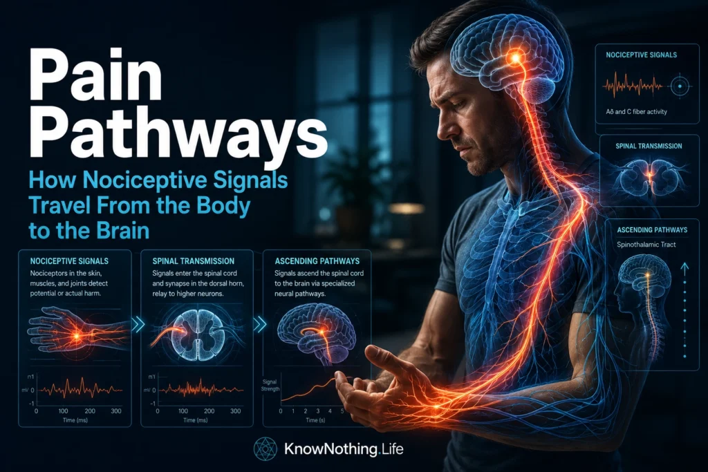
Pain pathways are the neural routes through which potentially damaging events are detected, transmitted, modified, and transformed into conscious pain. The familiar diagram of a signal traveling from an injured hand to the brain is useful but incomplete. Nociceptive information moves through several parallel pathways, encounters processing at every level, and is continuously influenced by inflammation, attention, expectation, emotion, memory, and descending signals from the brain. Pain is therefore not a passive message carried along a single biological wire. It is an active protective response generated by interacting peripheral, spinal, brainstem, thalamic, and cortical systems.
Nociception and pain are related but not identical. Nociception refers to the neural encoding of actual or potential tissue threat, whereas pain is the conscious sensory and emotional experience that may follow. Protective spinal reflexes can occur before conscious awareness, and nociceptive activity can be amplified or suppressed before it reaches the cortex. Ronald Melzack and Patrick Wall’s landmark 1965 paper “Pain Mechanisms: A New Theory” challenged the simple idea that pain intensity must correspond directly to injury. Their gate control theory proposed that spinal circuits and descending brain activity actively regulate nociceptive transmission.
Peripheral Detection of Threat
Pain pathways often begin at the free nerve endings of nociceptors located in the skin, joints, muscles, blood vessels, bones, and internal organs. These sensory neurons contain molecular receptors and ion channels that respond to potentially damaging heat, cold, pressure, acidity, inflammation, and irritating chemicals. When activated, the channels allow charged particles to enter the neuron, changing its electrical state and potentially initiating an action potential. Different populations of nociceptors are specialized for different forms of threat, although many respond to combinations of mechanical, thermal, and chemical stimulation.
A major advance came in 1997 when Michael Caterina, David Julius, and colleagues cloned the capsaicin receptor, now known as TRPV1. They demonstrated that this ion channel could be activated both by capsaicin—the ingredient responsible for the burning sensation of chili peppers—and by noxious heat. The discovery provided a molecular explanation for how certain environmental stimuli are converted into electrical activity within sensory neurons. Experiments in mice lacking TRPV1 later showed impaired responses to painful heat and reduced inflammatory thermal hypersensitivity, confirming that the receptor performs an important function within peripheral nociceptive pathways.
Carrying Signals Toward the Spinal Cord
Once a nociceptor is activated, electrical impulses travel along its axon toward the spinal cord. Thinly myelinated A-delta fibers conduct signals relatively quickly and are commonly associated with sharp, immediate, reasonably well-localized pain. Unmyelinated C fibers conduct more slowly and frequently contribute to burning, aching, throbbing, or diffuse sensations that continue after the first sharp response. These categories are not absolute, but they help explain why an injury can produce an immediate sting followed by a slower and more persistent discomfort.
Voltage-gated sodium channels allow these impulses to propagate along peripheral nerves. The importance of one such channel, Nav1.7, became clear through research on families whose members could not experience pain normally. In 2006, James Cox and colleagues identified loss-of-function mutations in the SCN9A gene, which encodes Nav1.7, in people with congenital inability to experience pain. Other forms of sensation remained largely intact, but injuries, burns, and fractures failed to produce ordinary pain. The findings provided direct human evidence that peripheral nerve excitability is essential to the functioning of normal pain pathways.
Processing Within the Spinal Cord
The cell bodies of primary nociceptive neurons are located in dorsal root ganglia beside the spinal cord. Sensory signals from much of the face instead enter through the trigeminal system. Within the spinal cord, primary afferent fibers terminate mainly in the dorsal horn, where they release glutamate and neuropeptides onto second-order neurons and interneurons. The dorsal horn is not merely a junction through which information passes unchanged. Local excitatory and inhibitory circuits compare nociceptive signals with touch, movement, and commands descending from the brain.
Gate control theory proposed that activity from larger touch-sensitive fibers could activate inhibitory spinal mechanisms and reduce the transmission of nociceptive input. This helps explain why rubbing or pressing near an injury can temporarily lessen discomfort. The theory’s original circuitry has been revised as scientists have identified additional neuron types, receptors, and pathways, but its central principle remains influential: nociceptive information is filtered and regulated within the spinal cord before it contributes to conscious pain.
Strong or repeated nociceptive input can also change how dorsal-horn neurons respond. In a landmark 1983 experiment, Clifford Woolf showed that intense peripheral stimulation could produce prolonged increases in the excitability of spinal neurons. Woolf and Stephen Thompson later demonstrated that NMDA receptor activity was important for establishing and maintaining this hypersensitive condition. Known as central sensitization, the process can cause painful stimuli to feel more intense, expand the area of tenderness, and allow ordinarily harmless touch to produce pain.
Ascending Pain Pathways
After spinal processing, many second-order neurons cross to the opposite side of the spinal cord and ascend through the anterolateral system. Its best-known component is the spinothalamic tract, which carries information concerning noxious stimulation, temperature, and certain forms of crude touch toward the brainstem and thalamus. Human research using laser-evoked potentials has measured conduction through the spinothalamic system by selectively activating small sensory fibers, helping clinicians assess pathway function in people with spinal cord disorders.
The anterolateral system is not one uninterrupted cable. Spinoreticular projections reach brainstem networks involved in arousal and defensive behavior, while spinomesencephalic fibers communicate with the midbrain periaqueductal gray. Other spinal neurons project to the parabrachial nucleus, which connects with the amygdala, hypothalamus, and related structures. Animal studies have shown that the parabrachial-to-amygdala circuit contributes to aversion, threat learning, escape behavior, and injury-induced pain sensitization. These parallel routes help explain why pain can simultaneously produce a localized sensation, fear, vigilance, autonomic changes, and an urgent motivation to protect the body.
Visceral pain also demonstrates the complexity of ascending transmission. Internal organs are often represented less precisely than the skin, and visceral and somatic inputs may converge on some of the same spinal neurons. This convergence can contribute to referred pain, in which an internal problem is experienced in a distant area of the body. Research on primate spinothalamic neurons has shown that cells receiving visceral input may also respond to stimulation of skin, muscles, or other somatic structures from related spinal segments.
From the Thalamus to the Cortex
The thalamus distributes nociceptive information to several cortical and subcortical regions rather than sending it to a single pain center. Lateral thalamic pathways communicate with somatosensory areas that contribute to identifying the location, duration, and intensity of stimulation. Other thalamic nuclei project toward the insula, anterior cingulate cortex, and limbic networks involved in bodily awareness, unpleasantness, motivation, and emotional significance. Pain therefore emerges from distributed processing rather than from activity in one specialized anatomical structure.
The posterior insula appears to contribute to representing the location and physical qualities of potentially damaging stimulation. In a high-resolution functional imaging study, Jennifer Brooks and colleagues delivered painful heat to the face, hand, and foot and observed an organized bodily map within the dorsal posterior insula. The finding supported the idea that the insula receives structured information about the physiological condition of the body rather than contributing only to generalized emotional distress.
Other regions add cognitive, emotional, and motivational dimensions. The anterior cingulate cortex participates in unpleasantness and action selection, while prefrontal regions help evaluate meaning, expectation, context, and possible responses. The amygdala contributes to threat learning and emotional salience. In 2013, Tor Wager and colleagues identified a multiregional functional MRI pattern that predicted experimentally induced heat pain across participants and distinguished it from several nonpainful states. The study showed that pain has measurable systems-level signatures, while also confirming that no individual region functions as a universal pain meter.
Descending Pain Modulation
Pain signals do not travel only upward. The cerebral cortex, hypothalamus, amygdala, and anterior cingulate cortex communicate with brainstem centers that send signals back toward the spinal dorsal horn. One major descending circuit passes through the periaqueductal gray and the rostral ventromedial medulla. These structures can recruit endogenous opioid, serotonin, noradrenaline, and other signaling systems that alter the responsiveness of spinal neurons and regulate how much nociceptive information continues upward.
Classic experiments showed that electrical stimulation or morphine administration within the periaqueductal gray could produce powerful analgesia through a descending pathway involving the medulla. In 1979, M. M. Behbehani and Howard Fields demonstrated an excitatory connection from the periaqueductal gray to the nucleus raphe magnus, helping establish the anatomy of this regulatory circuit. Descending systems can suppress pain when survival requires continued action, but they can also facilitate spinal transmission and increase sensitivity under other circumstances.
This bidirectional control helps explain why attention, expectation, fear, mood, sleep, and prior experience can alter pain without changing the original injury. These factors do not make pain imaginary. They change biological activity within pathways that regulate nociceptive processing. The nervous system continually evaluates whether pain should be intensified to encourage protection, reduced to permit urgent action, or maintained to prevent further harm.
When Pain Pathways Become Altered
Inflammation, nerve injury, or prolonged nociceptive input can produce plastic changes throughout the pain system. Peripheral receptors may become easier to activate, injured nerves may fire spontaneously, spinal inhibition may weaken, and central neurons may respond excessively. Signals that previously represented normal touch can then gain access to sensitized circuits, producing allodynia. Persistent pain may also involve altered thalamic, limbic, and descending activity, meaning that the mechanisms maintaining pain can eventually differ from the event that originally started it.
These distinctions have practical consequences. Nociceptive pain driven primarily by inflammation may respond to treatments that reduce peripheral signaling. Neuropathic pain caused by a lesion or disease of the somatosensory system may require medications or stimulation techniques aimed at abnormal neural excitability. Local anesthetics interrupt peripheral conduction, spinal cord stimulation modifies activity in spinal and ascending networks, and rehabilitative or psychological interventions can influence movement, threat evaluation, attention, and descending modulation. No single treatment targets every stage of the pathway.
Pain-pathway research increasingly combines genetics, electrophysiology, quantitative sensory testing, imaging, and computational analysis. Scientists are mapping specialized sensory-neuron populations, examining communication between the nervous and immune systems, and developing closed-loop stimulation that responds to changing neural activity. The central lesson is already clear: the route from tissue to experience is neither straight nor fixed. It is a multilayered biological system that detects danger, filters information, assigns meaning, organizes protective action, and changes through experience.
Understanding these pathways prevents two opposite errors. Pain should not be treated as a perfect measurement of tissue damage, but it should not be dismissed when scans fail to reveal an injury proportional to the symptoms. Every pain experience is produced through biological pathways, even when the most important changes involve sensitization, modulation, or altered processing rather than continuing tissue damage. By tracing nociception from peripheral receptors to the spinal cord, brain, and descending systems, neuroscience provides a stronger foundation for diagnosis, treatment, and respect for the person experiencing pain.


