
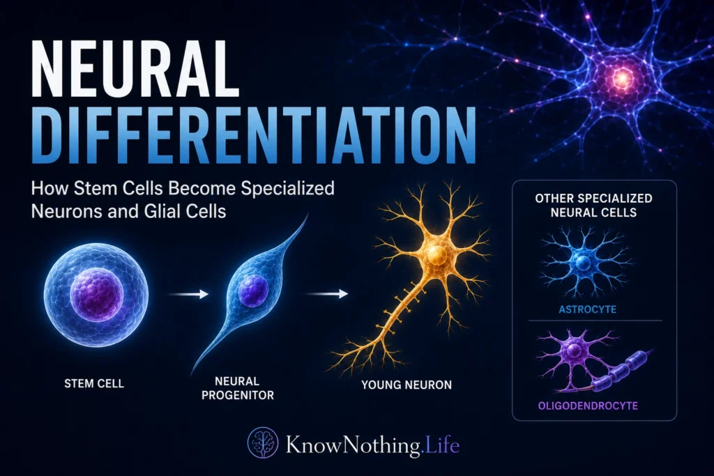
Neural differentiation is the process through which immature cells acquire the specialized identities and functions of the nervous system. During early development, embryonic cells first become committed to a neural fate and then give rise to neural stem and progenitor cells. These progenitors can divide, maintain an undifferentiated population, or produce neurons, astrocytes, and oligodendrocytes. Differentiation changes nearly every aspect of a cell, including its shape, gene expression, electrical properties, metabolism, connections, and responsiveness to surrounding signals.
The process is not a simple sequence in which one master gene turns a generic cell into a mature neuron. Cell fate develops through interactions among extracellular signals, transcription factors, epigenetic mechanisms, cell-cycle regulators, and neighboring cells. A progenitor’s location and developmental history influence which signals it can interpret, while the timing and intensity of those signals help determine whether it becomes a neuron or glial cell. Neural differentiation therefore combines inherited developmental programs with information supplied by the cell’s local environment.
Neural Induction and the Beginning of Neural Identity
Before cells can differentiate into particular neural types, part of the embryonic ectoderm must become neural tissue. Experiments in Xenopus embryos helped reveal how this initial neural induction occurs. In 1995, Ali Hemmati-Brivanlou and colleagues reported that bone morphogenetic protein 4, or BMP4, promotes epidermal development while inhibiting neural fate. Other studies showed that organizer-produced proteins such as Chordin and Noggin antagonize BMP signaling, allowing ectodermal cells to express neural markers instead of becoming epidermis. These findings contributed to the “default model,” according to which suppression of BMP activity permits an underlying neural developmental program to emerge.
BMP inhibition is essential, but later research showed that neural induction cannot always be explained by one signal alone. Experiments in chick and amphibian embryos found that fibroblast growth factor signaling, Wnt regulation, and other pathways can cooperate with BMP antagonism to establish and stabilize neural identity. Neural induction is therefore better understood as a coordinated transition in which cells lose responsiveness to epidermal instructions while activating transcriptional programs that make neural development possible.
Maintaining Progenitors or Producing Neurons
The developing nervous system must balance two competing demands. It must generate differentiated cells, but it must also preserve enough progenitors to continue growth. If every neural stem cell differentiated at once, the progenitor pool would be exhausted before the brain and spinal cord were fully formed. Signaling through the Notch pathway helps maintain this balance by discouraging premature neuronal differentiation and preserving cells in an undifferentiated or slowly dividing state.
Notch often operates through lateral inhibition. A cell beginning to express proneural genes can increase production of Delta ligands, which activate Notch receptors on neighboring cells. Notch activation induces proteins such as HES1 that suppress proneural gene expression, making nearby cells less likely to differentiate at the same moment. Studies in vertebrate embryos found that Delta–Notch signaling preserves populations of uncommitted neural cells, while genetic disruption of the pathway produces excessive or mistimed neuronal differentiation. Mouse experiments have also shown that Notch signaling is required to maintain neural stem-cell populations in several embryonic brain regions.
Notch activity is dynamic rather than permanently high or low. HES1 and proneural genes can oscillate within progenitor cells, allowing cells to remain responsive while delaying irreversible commitment. When proneural expression becomes sustained and Notch inhibition is overcome, the cell is more likely to leave the cell cycle and begin neuronal differentiation. This timing mechanism helps produce neurons gradually while retaining progenitors for later developmental stages.
Morphogens and Regional Cell Identity
A cell must become not only neural but also the correct kind of neural cell for its position. The nervous system contains motor neurons, sensory neurons, interneurons, dopamine-producing neurons, astrocytes, oligodendrocytes, and many other specialized populations. Secreted signaling molecules called morphogens provide positional information by forming concentration gradients across developing tissue. Cells exposed to different concentrations or durations of a morphogen activate different combinations of transcription factors.
Sonic Hedgehog is one of the best-understood examples. It is released from structures near the ventral neural tube and organizes the differentiation of several ventral neuronal populations. In a landmark study, Johan Ericson and colleagues showed that Sonic Hedgehog could induce distinct ventral neural cell types along much of the developing central nervous system. Later research demonstrated that Sonic Hedgehog controls differentiation through graded activity of GLI transcription factors and that both signal concentration and exposure time contribute to the identity adopted by a progenitor.
Morphogens do not act as simple labels. Sonic Hedgehog can influence patterning, proliferation, and survival, while BMPs, Wnts, retinoic acid, and fibroblast growth factors contribute additional spatial information. A cell’s response depends on its previous exposure, receptor expression, chromatin state, and interactions with other pathways. Neural identity emerges from combinations of signals interpreted through gene-regulatory networks rather than from one molecule acting alone.
Proneural Genes and Neuronal Commitment
Proneural transcription factors convert positional and developmental information into a neuronal program. Many belong to the basic helix-loop-helix family, including ASCL1, formerly called MASH1, and the neurogenins. These factors activate genes that promote cell-cycle exit, neuronal structure, migration, and electrical specialization. François Guillemot and colleagues found that ASCL1 and NEUROG1 are expressed in complementary regions of the developing nervous system, helping define progenitor populations that later produce functionally different neurons.
Proneural factors perform both general and subtype-specific functions. They encourage progenitors to become neurons, but they can also help determine what kind of neuron develops. Gene-replacement experiments showed that ASCL1 and NEUROG2 are not always interchangeable: their ability to produce a general neuronal fate can overlap, while their capacity to specify regional neuronal identities differs. NEUROG2, for example, is required for important stages in the differentiation of ventral midbrain dopamine neurons.
NeuroD acts later in many neuronal lineages and helps drive terminal differentiation. In a classic 1995 experiment, ectopic NeuroD expression converted broad areas of Xenopus embryonic ectoderm into neurons and caused premature differentiation of neural precursors. The study demonstrated that a transcription factor could activate a substantial portion of the neuronal program even in cells that would not normally differentiate at that time or location.
The Transition From Neurogenesis to Gliogenesis
Neural progenitors do not produce all cell types simultaneously. In much of the central nervous system, neurons are generated before most astrocytes and oligodendrocytes. This sequence allows early neurons to establish circuits while progenitor cells later change their competence and begin producing glia. The transition depends on signaling pathways, transcription factors, and changes in chromatin that make glial genes increasingly accessible.
SOX9 is a major regulator of glial differentiation. In mice lacking Sox9 in neural progenitors, the developing spinal cord continued producing neurons instead of making the normal transition toward astrocytes and oligodendrocytes. These results showed that SOX9 helps end the strongly neurogenic phase and establishes glial competence. Notch signaling can also promote astroglial differentiation partly through SOX9, illustrating how the same pathway can preserve early progenitors and influence later cell-fate choices depending on developmental context.
Epigenetic regulation contributes to this changing competence. DNA methylation initially restricts access to several astrocyte-related genes. A study led by Guo-li Fan found that loss of the maintenance methyltransferase DNMT1 caused premature activation of JAK–STAT signaling and early astrocyte differentiation. More recent experiments showed that TET-dependent DNA demethylation is particularly important for establishing the ability of neural stem cells to produce astrocytes and oligodendrocytes, even when neuronal differentiation remains possible.
Differentiation Is Not the Same as Maturation
A newly differentiated neuron is not yet a fully functional component of a neural circuit. It must extend an axon and dendrites, acquire appropriate neurotransmitters and receptors, develop voltage-gated ion channels, form synapses, and integrate incoming activity. Survival often depends on neurotrophic support and successful connections with target cells. Electrical activity then helps refine synapses, stabilize useful pathways, and eliminate inappropriate connections.
The distinction between differentiation and maturation is especially important in laboratory models. Human neurons derived from induced pluripotent stem cells may express neuronal markers before they display mature electrical and synaptic properties. Electrophysiological studies have shown that sodium and potassium currents develop in a coordinated manner over weeks as stem-cell-derived neurons become capable of producing repetitive action potentials. Other research has demonstrated that these cells can form identifiable pre- and postsynaptic structures, making them useful for studying human synaptic development and neurological disease.
Stem-Cell Models and Regenerative Medicine
Human embryonic stem cells and induced pluripotent stem cells have made it possible to study neural differentiation in living human cells. Researchers can expose pluripotent cells to combinations of signaling molecules that mimic embryonic development, producing neural progenitors and then directing them toward cortical, spinal, dopaminergic, or glial identities. Patient-derived cells can preserve disease-relevant genetic variation, allowing scientists to investigate conditions that cannot be studied adequately in postmortem tissue or animal models.
Transcription-factor-based methods can also accelerate differentiation. Forced expression of NGN2 can convert human pluripotent stem cells into functional neurons far more rapidly than many traditional developmental protocols. However, speed creates limitations: rapidly induced neurons may bypass intermediate stages and may not reproduce every aspect of normal regional development. Neural differentiation models are therefore powerful tools for disease research, drug testing, and regenerative medicine, but their cellular identity and maturity must be evaluated carefully.
Neural differentiation reveals how cells with essentially the same genome can become profoundly different biological structures. A neuron, astrocyte, and oligodendrocyte use different sets of genes because developmental signals and regulatory mechanisms open some pathways while closing others. The resulting identity is not created in one moment. It is constructed through successive decisions that connect embryonic position, cell-to-cell communication, transcription, epigenetic regulation, and activity into a stable neural fate.


