
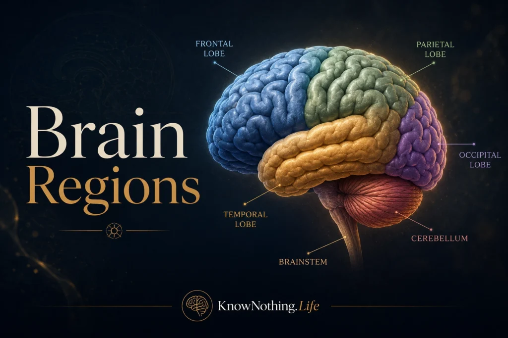
The brain is often described as the most complex organ in the human body, but that complexity is not random. It is organized into regions, systems, pathways, and networks that support sensation, movement, memory, emotion, language, attention, decision-making, and consciousness. Brain regions are not little machines working alone. They are specialized areas that communicate constantly, shaping the unified experience of being alive. To study brain regions is to study how physical tissue becomes perception, thought, feeling, action, and identity.
For centuries, philosophers and physicians wondered whether mental functions were spread evenly through the brain or localized in specific places. Modern neuroscience shows that both ideas are partly true. Certain regions are highly specialized: the occipital lobe is central to vision, the hippocampus is crucial for memory, and the motor cortex helps control voluntary movement. Yet no complex human function belongs entirely to one spot. Language, self-control, emotion, imagination, and awareness emerge from networks. The brain is regional, but it is also relational.
The Cerebral Cortex
The cerebral cortex is the folded outer layer of the brain and one of the most important structures for higher mental life. Its wrinkles, called gyri and sulci, increase surface area, allowing more neural tissue to fit inside the skull. The cortex is divided into two hemispheres, left and right, connected by the corpus callosum. Although the hemispheres are often simplified as “logical left” and “creative right,” real brain lateralization is much more complex. Both hemispheres contribute to thought, movement, emotion, and perception.
Neuroanatomists often divide the cortex into four major lobes: frontal, parietal, temporal, and occipital. Some also identify the insula and limbic cortex as distinct regions. These divisions are useful because they describe broad functional territories. The frontal lobe is strongly linked with planning and action. The parietal lobe integrates sensory and spatial information. The temporal lobe supports hearing, memory, and meaning. The occipital lobe processes vision. Still, each lobe contains many subregions, and nearly every important behavior depends on cooperation among several areas.
The Frontal Lobe
The frontal lobe sits behind the forehead and is central to planning, judgment, voluntary movement, attention, impulse control, and personality. The primary motor cortex, located near the back of the frontal lobe, sends commands that help control voluntary movement. Wilder Penfield’s famous brain stimulation studies helped reveal a motor “homunculus,” a map showing how different body parts are represented in the cortex. Hands, lips, and face occupy large cortical areas because they require fine control.
The prefrontal cortex is especially important for executive function. It helps people plan, delay gratification, shift attention, evaluate consequences, regulate emotion, and choose goals. Damage to frontal regions can produce impulsivity, poor judgment, emotional changes, apathy, or difficulty organizing behavior. The famous case of Phineas Gage, a railroad worker who survived a severe frontal brain injury in 1848, became one of the early examples suggesting that personality and moral judgment are connected to brain structure. The frontal lobe does not create character alone, but it helps organize the self in action.
The Parietal Lobe
The parietal lobe is located near the upper back portion of the brain and is essential for touch, body awareness, spatial processing, attention, and sensory integration. The primary somatosensory cortex receives information from the body, including pressure, temperature, pain, and position. Like the motor cortex, it contains a body map. Areas with high sensitivity, such as the hands, lips, and face, receive larger representation.
Beyond touch, the parietal lobe helps the brain understand where the body is in space. It allows people to reach for objects, judge distance, navigate rooms, and coordinate vision with movement. Damage to the right parietal lobe can produce hemispatial neglect, a condition in which a person may ignore the left side of space, even though their eyes still function. This shows that perception is not simply receiving visual input. The brain must organize attention and spatial meaning. The parietal lobe helps construct the body’s relationship to the world.
The Temporal Lobe
The temporal lobe lies along the side of the brain near the ears and is deeply involved in hearing, language comprehension, memory, emotion, and object recognition. The primary auditory cortex processes sound, while surrounding areas help interpret speech, music, tone, and complex auditory patterns. In the left temporal lobe, Wernicke’s area is strongly associated with language comprehension. Damage there can produce fluent but meaningless speech and difficulty understanding spoken language.
The temporal lobe also contains the hippocampus, one of the most important structures for forming new memories. The case of Henry Molaison, known as H.M., transformed neuroscience after surgery removed parts of his medial temporal lobes to treat epilepsy. Afterward, he could no longer form many new long-term declarative memories, though his intelligence and some forms of learning remained. This revealed that memory is not a single ability and that the medial temporal lobe is essential for turning experience into lasting memory.
The Occipital Lobe
The occipital lobe, located at the back of the brain, is the primary center for visual processing. Information from the eyes travels through the optic nerves, thalamus, and then to the primary visual cortex. From there, visual information moves through pathways that help identify objects, detect motion, analyze color, judge depth, and guide action. Vision feels effortless, but it is one of the brain’s most complex achievements.
Damage to the occipital lobe can produce blindness in parts of the visual field, even when the eyes are healthy. Some people with damage to visual areas experience visual agnosia, where they can see objects but cannot recognize them. Others may retain unconscious visual abilities despite reporting blindness, a phenomenon known as blindsight. These conditions show that seeing is not merely a matter of light entering the eye. The brain must interpret, organize, and give meaning to visual information.
The Limbic System
The limbic system is a network of structures involved in emotion, memory, motivation, threat detection, and bodily regulation. Major limbic structures include the amygdala, hippocampus, hypothalamus, cingulate cortex, and parts of the thalamus. The term “limbic system” became influential through researchers such as Paul MacLean, though modern neuroscience treats it less as a single emotional brain and more as a set of interacting networks.
The amygdala helps detect emotional significance, especially threat and fear. It contributes to emotional learning, helping the brain remember what is dangerous or important. The hippocampus supports memory formation and context. The hypothalamus regulates hunger, thirst, sleep, temperature, hormones, and stress responses. The cingulate cortex helps connect emotion, attention, pain, and decision-making. Together, these structures show that emotion is not separate from cognition. Feeling, memory, body state, and attention are deeply intertwined.
The Basal Ganglia
The basal ganglia are deep brain structures involved in movement, habit, reward, motivation, and action selection. They include the caudate nucleus, putamen, globus pallidus, substantia nigra, and related circuits. These structures help the brain initiate desired actions and inhibit unwanted ones. They are essential for smooth movement, procedural learning, and the formation of habits.
Disorders of the basal ganglia reveal their importance. Parkinson’s disease involves degeneration of dopamine-producing neurons in the substantia nigra, leading to tremor, stiffness, slowed movement, and balance problems. Huntington’s disease affects basal ganglia circuits and can produce involuntary movement, emotional changes, and cognitive decline. Addiction and compulsive behavior also involve reward circuits connected to the basal ganglia. This region is not only about movement. It helps decide which actions become reinforced and repeated.
The Thalamus
The thalamus is often called the brain’s relay station, but it does more than pass messages along. Located deep in the brain, it routes sensory and motor signals to the cortex and helps regulate attention, awareness, and sleep-wake states. Nearly all sensory information except smell passes through thalamic circuits before reaching the cortex. The thalamus helps determine what information becomes available for higher processing.
Because of its central position, the thalamus plays an important role in consciousness. Damage to thalamic regions can affect alertness, perception, movement, and cognition. The thalamus works closely with the cortex in loops that help sustain attention and coordinated brain activity. It is not the “seat of consciousness,” but it is part of the architecture that allows conscious experience to remain organized and awake.
The Cerebellum
The cerebellum sits at the back and bottom of the brain and is traditionally associated with balance, coordination, timing, and motor learning. It helps movements become smooth, accurate, and adaptive. When the cerebellum is damaged, a person may have poor coordination, tremor during movement, unsteady walking, or difficulty with precise timing.
Modern research has expanded the cerebellum’s importance beyond movement. It appears to contribute to prediction, error correction, language, attention, and even emotional regulation. The cerebellum helps compare intended outcomes with actual results, allowing the brain to adjust. This may apply not only to physical movement but also to thought and behavior. Its dense circuitry makes it one of the brain’s most remarkable structures.
The Brainstem
The brainstem connects the brain to the spinal cord and includes the midbrain, pons, and medulla. It controls many basic life functions, including breathing, heart rate, swallowing, arousal, sleep cycles, and reflexes. The brainstem contains pathways that carry information between the brain and body, as well as nuclei connected to the cranial nerves.
The brainstem may not sound glamorous compared with the cerebral cortex, but it is essential for survival. Damage to the brainstem can be catastrophic because it supports the basic conditions that allow consciousness and bodily life to continue. The reticular formation, a network within the brainstem, helps regulate wakefulness and alertness. Before higher thought can occur, the organism must be awake, breathing, and physiologically stable. The brainstem helps make that possible.
Brain Regions Work as Networks
Although brain regions are useful to name and study, the brain works through networks. Language involves frontal, temporal, parietal, motor, auditory, and memory systems. Emotion involves the amygdala, prefrontal cortex, hippocampus, hypothalamus, body signals, and social interpretation. Decision-making requires reward circuits, memory, attention, impulse control, and prediction. Even a simple act like picking up a cup involves vision, spatial awareness, motor planning, touch, balance, and expectation.
This network view is one of the most important developments in modern neuroscience. The brain is not a collection of isolated boxes labeled “memory,” “emotion,” “language,” and “reason.” It is a living system of specialized regions communicating dynamically. Brain regions matter because specialization matters. But connection matters just as much. The mind arises not from one region alone, but from the organized activity of many regions working together.
Final Thoughts
Brain regions provide a map of the nervous system’s functional architecture. The frontal lobe supports planning, movement, and self-control. The parietal lobe integrates touch and space. The temporal lobe supports hearing, memory, and meaning. The occipital lobe processes vision. The limbic system links emotion and memory. The basal ganglia shape movement and habit. The thalamus routes and regulates information. The cerebellum refines coordination and prediction. The brainstem sustains life itself.
To understand brain regions is to understand that the mind is embodied. Thoughts, feelings, memories, decisions, and perceptions are not floating abstractions. They depend on tissue, pathways, chemistry, and electrical activity. Yet the brain is more than a set of parts. Its regions become meaningful through connection. Human experience emerges when specialized systems cooperate, compete, regulate, and integrate. The study of brain regions therefore reveals one of the deepest truths of neuroscience: the mind has a geography, but it lives through networks.


