
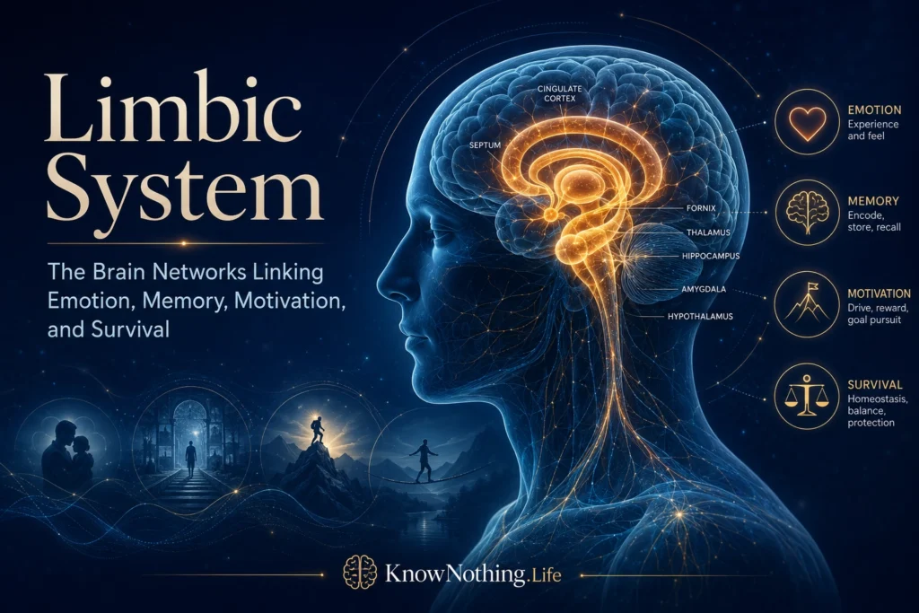
The limbic system is a widely used name for interconnected brain structures involved in emotion, memory, motivation, learning, bodily regulation, and adaptive behavior. It commonly includes the amygdala, hippocampal formation, cingulate cortex, hypothalamus, septal region, mammillary bodies, parts of the thalamus, and portions of the orbitofrontal and medial prefrontal cortex. These structures do not form a sealed anatomical organ with universally accepted borders. Instead, they participate in overlapping circuits that connect sensory information and remembered experience with autonomic reactions, hormonal responses, attention, decision-making, and action.
Popular descriptions sometimes call the limbic system the brain’s “emotional center,” but this phrase can be misleading. Emotions are not manufactured by one isolated collection of ancient structures while the cerebral cortex performs purely rational work. Emotional and cognitive functions continually influence one another, and many regions labeled limbic also contribute to spatial navigation, episodic memory, attention, social judgment, and planning. The term remains useful as a historical and educational framework, provided it is treated as shorthand for interacting networks rather than a single control center.
From the Papez Circuit to MacLean’s Model
The modern idea began with neuroanatomist James Papez, whose 1937 paper “A Proposed Mechanism of Emotion” described a circuit connecting the hippocampal formation, mammillary bodies of the hypothalamus, anterior thalamic nuclei, and cingulate gyrus. Papez proposed that the hypothalamus helped organize emotional expression, while activity reaching the cingulate cortex contributed to emotional experience. Although later research revised this interpretation, the Papez circuit established a lasting principle: emotion depends on communication among connected regions rather than activity in one location.
Paul MacLean expanded the proposal during the mid-twentieth century. In his 1952 paper on the “visceral brain,” he incorporated the amygdala, septal areas, and additional cortical and subcortical structures into what he called the limbic system. MacLean later placed the system within his triune-brain theory, portraying it as an evolutionarily old mammalian layer positioned between a reptilian complex and the rational neocortex. That model strongly influenced psychology and public education, but modern evolutionary neuroscience rejects its neatly layered account. Mammalian brains did not evolve by simply stacking a new rational brain over older emotional and instinctive brains.
Anatomy Without a Fixed Border
One difficulty in defining the limbic system is that its proposed membership has changed repeatedly. The hippocampus and cingulate cortex were central to Papez’s original circuit, whereas MacLean gave the amygdala and visceral regulation greater prominence. Later accounts added structures such as the nucleus accumbens, orbitofrontal cortex, ventromedial prefrontal cortex, insula, and parts of the basal forebrain. Each addition reflected genuine anatomical connections, but it also made the system’s boundaries less precise. Researchers including Joseph LeDoux and Edmund Rolls have therefore argued that neuroscience should describe specific circuits for emotional learning, reward, memory, attachment, and motivation rather than assume that one unified limbic system performs all emotional functions.
The structures traditionally grouped under the label are nevertheless densely interconnected. The amygdala communicates with sensory cortex, hippocampus, hypothalamus, striatum, brainstem, and prefrontal regions. The hippocampus connects remembered events and environmental contexts with present situations. The hypothalamus coordinates autonomic, endocrine, and behavioral responses related to hunger, thirst, reproduction, defense, and stress. Cingulate and prefrontal regions help assess significance, monitor conflict, calculate value, and alter behavior as circumstances change. Their functions arise from patterns of communication extending far beyond any conventional limbic boundary.
The Amygdala and Emotional Significance
The amygdala is often called the brain’s fear center, but decades of research support a broader role. Amygdala circuits help organisms learn that certain cues predict danger or reward, direct attention toward important stimuli, influence memory, and coordinate physiological and behavioral responses. Joseph LeDoux’s research on fear conditioning demonstrated how sensory information can reach amygdala pathways and support learned defensive reactions. Studies involving human patients and neuroimaging have also shown amygdala involvement in emotional learning, attention, perception, social signals, and memory for emotionally significant events.
This wider view explains why amygdala activity should not be equated with the conscious feeling of fear. Defensive reactions can begin before a person has fully identified a threat, while the subjective experience of fear requires broader systems that represent bodily changes, context, memory, and meaning. People with amygdala lesions may show specific impairments in recognizing danger or acquiring conditioned responses without losing every emotional capacity. The amygdala is therefore better understood as a collection of nuclei that assigns biological significance and helps other systems determine what deserves attention and action.
The Hippocampus, Cingulate Cortex, and Emotional Memory
The hippocampal formation is essential for forming episodic memories and representing context. It helps distinguish where and when an event occurred, whether a present situation resembles a past one, and which memories are appropriate to retrieve. In emotional learning, this contextual function can determine whether a defensive response is expressed in a genuinely dangerous environment or generalized to a safe one. Research on contextual fear conditioning shows that emotional behavior depends not only on detecting a cue but also on reconstructing the surrounding situation in which that cue acquired meaning.
The amygdala and hippocampus make complementary contributions to emotional memory. The hippocampus represents events and contexts, while the amygdala helps prioritize information associated with arousal or biological importance. Their interaction can strengthen long-term memory for significant experiences, although vividness does not guarantee accuracy. The cingulate cortex also participates in these processes. Research reviewed by Alexander Shackman and colleagues found overlapping anterior midcingulate involvement in negative affect, pain, and cognitive control, demonstrating again that emotion and cognition are integrated rather than divided into separate brain territories.
Homeostasis, Motivation, and Reward
The hypothalamus links neural processing with the internal condition of the body. Its specialized nuclei help regulate temperature, energy balance, thirst, reproductive functions, circadian rhythms, stress hormones, and autonomic activity. Larry Swanson’s work on motivated behavior describes hypothalamic and brainstem pathways that organize coordinated behavioral, hormonal, and bodily responses. Hunger, defense, caregiving, and reproduction are not simple reflexes generated by one instinct center; they involve sensory cues, internal needs, memory, learned expectations, and opportunities in the environment.
Motivation and reward also depend on circuits extending through the ventral tegmental area, nucleus accumbens, ventral pallidum, amygdala, hippocampus, thalamus, and prefrontal cortex. Suzanne Haber and Brian Knutson described this reward architecture as a cortical-basal-ganglia network rather than a single pleasure pathway. Dopamine contributes to learning, incentive value, and goal pursuit, while orbitofrontal and ventromedial prefrontal regions help represent changing outcomes and guide choices. These systems allow emotional significance to influence which goals are pursued, how much effort is invested, and when behavior should change.
The Limbic System in Modern Neuroscience
Contemporary neuroscience increasingly replaces the image of an isolated limbic system with network models. In “A Network Model of the Emotional Brain,” neuroscientist Luiz Pessoa describes functionally integrated cortical and subcortical systems linking the amygdala, hippocampus, striatum, hypothalamus, thalamus, brainstem, and multiple cortical regions. In this account, emotion is inseparable from perception, cognition, motivation, and action. A region’s contribution depends on the task, the moment, and its pattern of communication with the rest of the brain.
This network perspective also improves the study of psychiatric and neurological disorders. Anxiety, depression, post-traumatic stress, addiction, epilepsy, dementia, and traumatic brain injury may involve structures traditionally described as limbic, but none can be explained by one malfunctioning emotional center. Symptoms emerge through altered learning, memory, reward processing, bodily regulation, attention, and cognitive control. Modern models accordingly distinguish several partially overlapping networks responsible for memory, emotional valuation, self-directed thought, and adaptive behavior.
The enduring value of the limbic-system concept is not that it provides a perfect anatomical map. Its value lies in emphasizing a fundamental truth about the nervous system: feeling, remembering, valuing, and acting are closely connected. Emotional responses are shaped by previous experiences, bodily needs, environmental context, anticipated outcomes, and conscious interpretation. The limbic system is therefore best understood not as a primitive emotional brain hidden beneath the rational cortex, but as a historical name for interconnected pathways through which the brain coordinates the body, experience, and behavior.


