
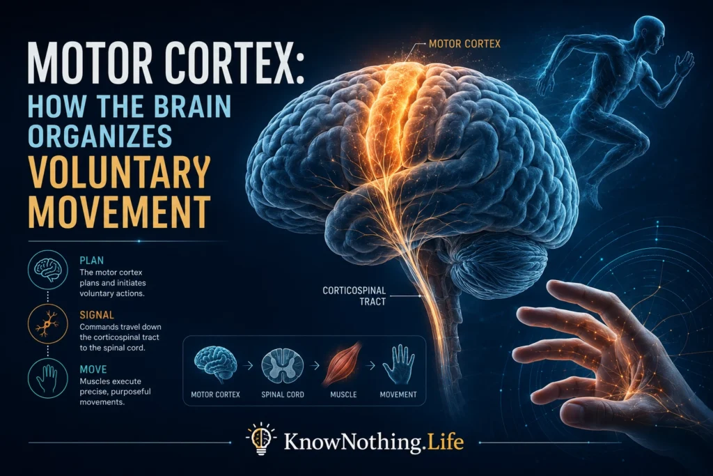
The motor cortex is a group of frontal-lobe regions that contributes to planning, initiating, and controlling voluntary movement. It includes the primary motor cortex, premotor cortex, supplementary motor area, and nearby cingulate motor regions. These areas exchange information with the parietal cortex, basal ganglia, cerebellum, thalamus, brainstem, spinal cord, and sensory systems. Even reaching for a cup requires the nervous system to identify a goal, estimate limb position, choose a trajectory, recruit suitable muscles, predict the result, and adjust the action as sensory information changes. Research on preparatory activity and population dynamics shows that motor control emerges from coordinated networks rather than from a single command center.
Primary motor cortex, commonly called M1, lies mainly along the precentral gyrus and provides an important source of descending influence over movement. Premotor regions help transform visual and other sensory information into actions, while the supplementary motor area contributes to internally generated movements, preparation, and coordination between the hands. These distinctions are useful, but their boundaries and functions overlap. Simultaneous recordings during visually guided grasping, for example, have revealed related but distinguishable contributions from primary and premotor cortex, while studies of supplementary motor activity have identified neurons preferentially active before self-initiated actions.
The Motor Homunculus and the Limits of a Simple Map
The best-known image of motor cortex is the motor homunculus, a distorted body mapped across the cortical surface. It grew from Wilder Penfield and Edwin Boldrey’s 1937 study, Somatic Motor and Sensory Representation in the Cerebral Cortex of Man as Studied by Electrical Stimulation. During neurosurgical procedures, stimulation of different cortical sites produced movements in different body regions. The face and hand occupied especially large territories, reflecting the complexity of their control rather than their physical size. The work established somatotopy—the orderly representation of body regions—as a major principle of motor-cortical organization.
Later research showed that the map is neither perfectly orderly nor divided into sharply separated body-part zones. Representations overlap, and one cortical location can influence several muscles or joints. In 2002, Michael Graziano, Charlotte Taylor, and Tirin Moore applied longer trains of microstimulation to monkey precentral cortex and evoked coordinated actions such as reaching, defensive movements, and bringing the hand toward the mouth. Their findings suggested that portions of motor and premotor cortex may be organized partly around useful actions or final postures, not merely isolated muscle contractions.
What Motor-Cortical Neurons Encode
Motor-cortical activity is related to many features of action, including force, direction, speed, posture, muscle activity, and behavioral context. Edward Evarts’s 1968 study, Relation of Pyramidal Tract Activity to Force Exerted During Voluntary Movement, showed that the firing of corticospinal neurons changed systematically with the force exerted by a monkey. The result challenged the view that motor cortex only specifies which muscles should contract and encouraged researchers to examine how neural activity reflects quantitative properties of movement. It also established recordings from awake, behaving animals as a powerful method for connecting neuronal discharge with voluntary action.
Apostolos Georgopoulos, Andrew Schwartz, and Ronald Kettner later demonstrated that individual motor-cortical neurons are broadly tuned to movement direction. In their 1986 paper, Neuronal Population Coding of Movement Direction, each neuron was represented as a directional vector weighted by its firing rate. The sum of the population vectors pointed toward the movement being performed. This model became a classic demonstration that precise motor information can emerge from the combined activity of many broadly tuned neurons rather than from one cell assigned to one movement.
Motor Cortex as a Dynamical System
Population coding does not mean that motor cortex holds a static description of an intended movement. Neural activity changes continuously before and during action, and these evolving patterns may help generate the commands needed to move. Mark Churchland and colleagues examined large neuronal populations during reaching and found orderly rotational patterns in neural state space. Their 2012 study, Neural Population Dynamics During Reaching, argued that motor-cortical activity can be understood partly as the evolution of a dynamical system rather than only as a representation of external variables such as direction or velocity.
This framework helps explain why the same neuron may appear related to different variables under different tasks. Its activity depends on the state of the wider network, the prepared action, and the patterns unfolding through time. Motor cortex can still contain information about direction, force, and muscle activity, but these variables may be useful descriptions of population dynamics rather than separate instructions encoded independently. The dynamical view therefore shifts attention from asking what one neuron represents to asking how coordinated neural trajectories generate structured muscular output.
Preparation Without Premature Movement
Motor-cortical activity often changes before movement begins. This preparatory activity creates an apparent problem: if motor cortex projects toward muscles, why does preparation not make the body move too soon? Matthew Kaufman and colleagues addressed this question in their 2014 study, Cortical Activity in the Null Space: Permitting Preparation Without Movement. They found that preparatory changes could occur in population patterns that had little effect on muscle-related output dimensions. When movement began, activity entered output-potent patterns capable of influencing downstream circuits.
The distinction between output-null and output-potent activity offers a mechanism through which motor networks can configure themselves before acting. Preparation may establish the initial neural state needed for a rapid and accurate trajectory after a go signal. Sensory cues, expectations, and task rules can influence this state through premotor and parietal networks. Motor preparation is therefore not simply a weaker version of execution. It is a structured computational stage that places the network in a condition from which the desired action can unfold without being released prematurely.
Corticospinal Output and Skilled Movement
Motor cortex influences movement through several descending routes, most prominently the corticospinal tract. Corticospinal axons descend through the internal capsule and brainstem, with most crossing near the junction between the medulla and spinal cord. They terminate on spinal interneurons and, particularly in primates, can also form relatively direct connections with motor neurons controlling hand and finger muscles. Anatomical and electrophysiological comparisons have shown that projections from M1 reach hand-muscle motor nuclei more densely than those from the supplementary motor area, helping explain M1’s importance for precise manual control.
The corticospinal tract is not a set of one-to-one cables linking individual cortical cells with individual muscles. Single descending neurons can influence several motor-unit populations, while each muscle receives converging input from overlapping cortical territories. Motor cortex also acts through corticobulbar, reticulospinal, and other pathways. Human lesions affecting motor cortex or the corticospinal tract can impair independent finger movements, producing weakness, poor fractionation, and reduced dexterity even when some gross or automatic actions remain possible.
Plasticity, Recovery, and Neural Interfaces
Motor-cortical organization changes with experience. Avi Karni and colleagues used functional MRI to study adults learning a rapid finger sequence and found changes in primary motor cortex associated with skill acquisition. Randolph Nudo and colleagues trained squirrel monkeys on a skilled hand task and reported expansion of cortical representations associated with the practiced movements. Comparable repetition without the same skill demands did not produce equivalent reorganization, indicating that meaningful learning can reshape motor maps more strongly than movement alone. This plasticity also contributes to recovery after injury, although compensatory changes are not always efficient.
Brain–computer interfaces make motor-cortical population signals visible in a practical way. Leigh Hochberg and colleagues showed that a person with tetraplegia could use activity recorded from motor cortex to control a computer cursor and external devices. The group later demonstrated that people with long-standing tetraplegia could guide a robotic arm through three-dimensional reaching and grasping, while Jennifer Collinger and colleagues reported high-performance prosthetic-arm control using two intracortical electrode arrays. These systems estimate intended movement from changing population patterns rather than reading one command neuron, reinforcing the central lesson of motor-cortex research: voluntary action emerges from distributed neural populations whose coordinated dynamics connect intention with movement.


