
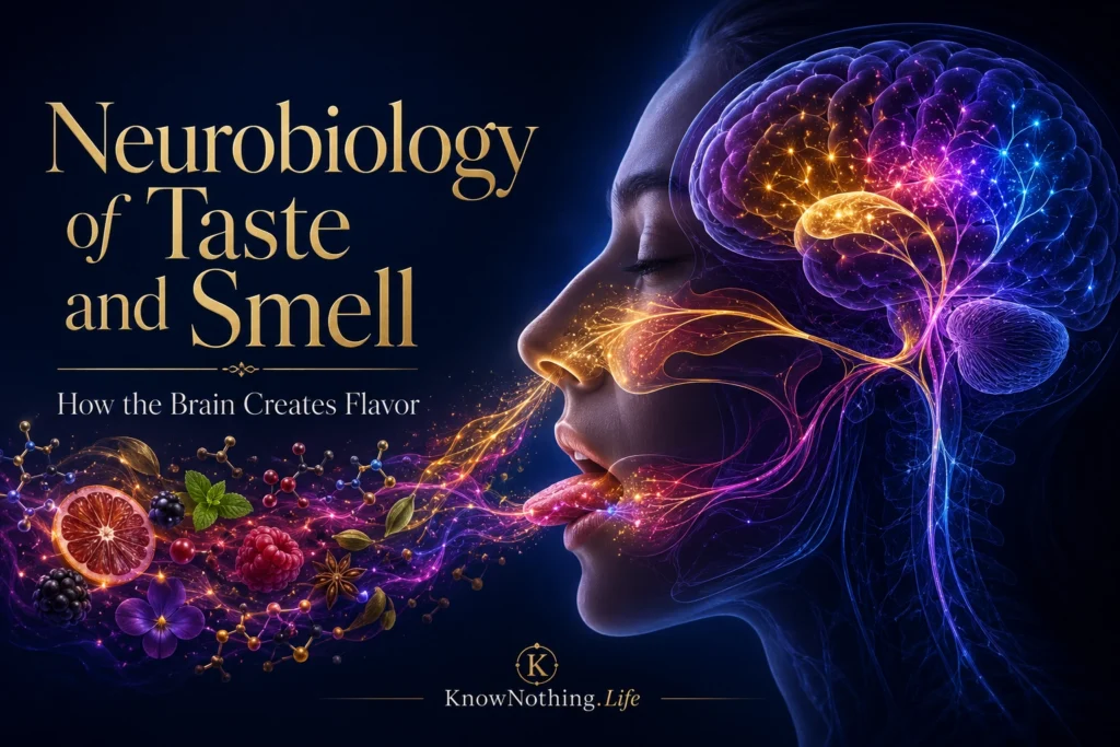
Taste and smell are the chemical senses, meaning that they begin when specialized receptors interact with molecules from food, beverages, the environment, or the body. Taste primarily evaluates substances dissolved in saliva, while smell detects volatile compounds entering the nasal cavity. Together, these systems help identify nutrients, avoid harmful substances, recognize familiar places and people, and regulate appetite. Their signals also combine with temperature, texture, vision, and oral sensations to produce flavor, a unified experience that cannot be explained by the tongue alone.
The two senses follow different anatomical routes but eventually converge in brain regions involved in perception, memory, emotion, and reward. Taste signals travel from the mouth through several cranial nerves to the brainstem, thalamus, insula, and frontal operculum. Olfactory signals travel from receptor neurons in the nose to the olfactory bulb and then reach areas including the piriform cortex, amygdala, entorhinal cortex, and orbitofrontal cortex. Their close relationship with emotional and motivational systems helps explain why smells and flavors can evoke vivid memories, change mood, or produce immediate attraction and disgust.
Taste Buds and Gustatory Receptor Cells
Taste begins in clusters of specialized epithelial cells called taste buds, located mainly within papillae on the tongue but also present on the soft palate, pharynx, and upper throat. Dissolved chemicals enter taste pores and contact receptor cells whose apical surfaces face the oral cavity. These cells are continually replaced and communicate with sensory fibers rather than sending axons directly into the brain. Different taste-cell populations detect sweet, bitter, umami, sour, and sodium-related stimuli through distinct receptors and ion channels.
Signals from the front of the tongue travel mainly through branches of the facial nerve, while the glossopharyngeal nerve carries information from the posterior tongue and the vagus nerve conveys input from parts of the throat. These fibers terminate in the rostral nucleus of the solitary tract in the brainstem. In primates, ascending information then reaches a gustatory region of the thalamus before projecting to the anterior insula and frontal operculum, which are frequently described as primary gustatory cortex. These pathways retain taste-quality information while also interacting with swallowing, digestion, and protective reflexes.
The Molecular Detection of Basic Tastes
Sweet, bitter, and umami tastes are detected largely through G-protein-coupled receptors. In 2001, Greg Nelson and colleagues demonstrated that T1R2 and T1R3 combine to form a receptor responsive to chemically diverse sweet substances, including sugars and artificial sweeteners. A related pairing of T1R1 and T1R3 functions as an amino-acid receptor associated with umami, the savory quality strongly elicited by glutamate. These receptors activate intracellular signaling pathways that change calcium levels and cause taste cells to release transmitter signals to nearby sensory nerve endings.
Bitter compounds are detected through the T2R receptor family. In 2000, Jayaram Chandrashekar and colleagues demonstrated that particular T2Rs respond to compounds such as cycloheximide, denatonium, and propylthiouracil. Bitter sensing is especially important because many toxic plant compounds produce bitter sensations, although bitterness does not always indicate toxicity. Sour taste depends strongly on the proton channel OTOP1, which allows acid-related signals to activate sour-responsive cells. Sodium attraction in mice depends partly on epithelial sodium channels, while concentrated salts also recruit aversive pathways. Human salt detection appears to involve multiple mechanisms rather than one fully defined receptor.
How Taste Cells Communicate with the Brain
Taste receptor cells must convert chemical detection into signals that sensory neurons can transmit. Cells detecting sweet, bitter, and umami compounds use a pathway involving intracellular calcium and release adenosine triphosphate, or ATP, as a neurotransmitter. Atsuko Taruno and colleagues showed in 2013 that the CALHM1 ion channel is essential for ATP-mediated transmission of these taste qualities. This finding revealed that ATP is not merely a cellular energy molecule in taste buds but also a signal carrying information from receptor cells to nerve fibers.
Taste coding continues to be debated in terms of labeled lines and population patterns. Peripheral receptor cells can be strongly specialized for particular qualities, and experiments manipulating receptor expression show that activating a sweet-cell pathway can produce attraction even when the activating molecule would not normally taste sweet. Higher brain regions, however, often contain overlapping populations that respond to several tastes, intensities, and internal states. Taste perception therefore appears to combine relatively selective peripheral channels with increasingly distributed cortical representations influenced by familiarity, hunger, expectation, and learned value.
Olfactory Receptors and the Nasal Epithelium
Smell begins in the olfactory epithelium high inside the nasal cavity. Olfactory sensory neurons extend cilia into a mucus layer where airborne molecules can bind to receptor proteins. Linda Buck and Richard Axel’s landmark 1991 paper, A Novel Multigene Family May Encode Odorant Receptors, identified a large family of genes encoding probable odorant receptors. This discovery provided the molecular foundation for understanding how the olfactory system can discriminate among an enormous variety of chemicals without requiring one receptor for every recognizable smell.
An odorant can activate several receptor types, and one receptor can respond to multiple odorants. Smell identity is therefore represented through combinations of receptor activity. Individual mature olfactory neurons generally express one receptor gene from the available repertoire. Neurons expressing the same receptor are scattered across zones of the nasal epithelium, but their axons converge onto a small number of structures called glomeruli within the olfactory bulb. Peter Mombaerts and colleagues visualized this organization genetically in 1996, revealing a receptor-specific sensory map at the brain’s entrance to the olfactory system.
From the Olfactory Bulb to Odor Perception
Within each glomerulus, olfactory sensory neurons communicate with projection neurons known as mitral and tufted cells. Interneurons surrounding these circuits regulate timing, contrast, and interactions among glomeruli. Odors consequently evoke patterns distributed across multiple glomeruli rather than activating one dedicated “coffee,” “rose,” or “smoke” location. These patterns also change with concentration and the timing of inhalation, requiring the nervous system to identify stable odor objects from fluctuating chemical input.
Mitral and tufted cells project to several brain regions without first passing through the same obligatory thalamic relay used by vision, hearing, and somatic sensation. One major destination is the piriform cortex. Research suggests that piriform representations are distributed across ensembles rather than arranged as a simple spatial map. Jay Gottfried and colleagues found that different portions of human piriform cortex carry information related to molecular structure and perceived odor quality. This transformation helps the brain recognize similar-smelling substances even when their chemical structures differ and distinguish chemicals that are structurally similar but smell different.
How Taste and Smell Become Flavor
Much of what people casually call taste is produced by retronasal olfaction. During chewing and swallowing, volatile molecules travel from the mouth through the back of the throat into the nasal cavity. The brain interprets this olfactory input as originating from food in the mouth rather than from the nose. When nasal airflow is blocked, sweet, salty, sour, bitter, and umami sensations remain, but the identities of coffee, chocolate, herbs, fruits, and other foods become far more difficult to distinguish.
Flavor also includes touch, temperature, and irritation. Trigeminal nerve endings detect qualities such as the burn of chili peppers, the cooling effect of menthol, carbonation, creaminess, and astringency. Taste, retronasal smell, and oral somatosensation converge in regions including the insula and orbitofrontal cortex. Dana Small and colleagues found that familiar taste-and-odor combinations produced enhanced responses in areas associated with flavor integration. Their work indicated that the brain’s merging of smell and taste is shaped by experience rather than being a purely automatic addition of two sensory signals.
Appetite, Reward, Memory, and Emotion
Taste and smell provide information about identity, but they also assign value. Sweetness may signal accessible carbohydrates, umami may indicate amino acids, and excessive bitterness or sourness may warn of potentially unsuitable food. These meanings are not fixed. Hunger can increase the attraction of food odors, while eating to satiety can reduce the pleasantness of the same flavor. The orbitofrontal cortex plays a particularly important role in representing pleasantness and reward value, integrating chemosensory information with current needs and previous outcomes.
Olfactory pathways also communicate closely with the amygdala and memory-related areas of the medial temporal lobe. This anatomy helps odors become associated with emotional events, places, illnesses, and social experiences. A smell paired with nausea can later trigger avoidance, while aromas associated with safety or family traditions may become strongly comforting. Learning can reorganize odor categories and flavor preferences, demonstrating that chemosensory perception reflects both molecular stimulation and acquired meaning.
Plasticity, Sensory Loss, and Future Research
Taste and olfactory systems are unusually dynamic. Taste receptor cells are regularly replaced, and the olfactory epithelium contains basal cells capable of producing new sensory neurons throughout adult life. Regeneration is not always complete, however. Viral illness, head injury, medications, aging, toxic exposure, neurodegenerative disease, and local inflammation can reduce taste or smell. Loss of smell can also be mistaken for loss of taste because the five basic taste qualities may remain while the richer olfactory components of flavor disappear.
Future research is combining genetics, single-cell analysis, brain imaging, neural recording, and computational modeling to clarify how chemical patterns become conscious qualities. Scientists are also investigating methods to regenerate receptor cells, restore olfactory connections, diagnose early neurological disease, and predict how receptor differences influence diet and health. The central challenge is to explain how separate signals for molecules, temperature, texture, memory, and bodily need are fused into one experience. Taste and smell begin with chemistry, but flavor is created by the coordinated activity of the brain.


