
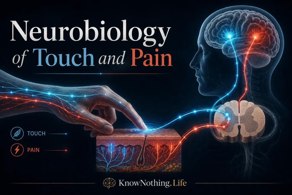
Touch and pain belong to the somatosensory system, the network through which the nervous system monitors the body and its physical surroundings. This system detects pressure, vibration, skin stretch, temperature, tissue damage, limb position, and movement. Although touch and pain often interact, they serve different biological functions. Touch supplies detailed information used to recognize objects, control grip, maintain posture, and communicate socially. Pain is a protective experience that motivates withdrawal, guarding, learning, and recovery when the nervous system judges that the body is threatened.
Neither sensation is a direct copy of physical stimulation. Mechanical pressure or heat must first be converted into electrical activity by sensory receptors. Those signals are then filtered and reorganized in peripheral nerves, the spinal cord, brainstem, thalamus, and cerebral cortex. Pain is especially dependent on interpretation: nociception refers to neural activity associated with potentially damaging events, while pain is the conscious sensory and emotional experience that may result. Attention, expectation, memory, stress, and context can therefore alter pain without changing the original injury.
Skin Receptors and the Neural Coding of Touch
The skin contains several classes of low-threshold mechanoreceptors that respond to ordinary physical contact. Merkel cell–neurite complexes are particularly sensitive to sustained pressure, edges, and spatial detail. Meissner corpuscles respond rapidly to skin movement and low-frequency vibration, while Pacinian corpuscles are exceptionally sensitive to brief changes and higher-frequency vibration. Ruffini-associated endings respond to sustained skin stretch. Recordings from individual human nerve fibers by Åke Vallbo and Roland Johansson showed that these receptor classes differ in receptive-field size, adaptation rate, and sensitivity, creating parallel information streams for shape, texture, vibration, slip, and hand movement.
Mechanical force must be translated into changes in neuronal voltage. A major part of this process depends on PIEZO2, a mechanically activated ion channel found in sensory neurons and specialized receptor cells. Sanjeev Ranade and colleagues showed that removing Piezo2 from mouse sensory neurons greatly reduced mechanically activated currents and impaired low-threshold touch responses. Human evidence came from Alexander Chesler and colleagues, who studied people carrying loss-of-function PIEZO2 variants. Participants had profound difficulties with vibration, discriminative touch, joint position, and coordinated movement, establishing PIEZO2 as a central molecular detector for human touch and proprioception.
Discriminative and Emotional Touch
Fast, detailed touch information is transmitted mainly through myelinated Aβ nerve fibers. These fibers enter the spinal cord and ascend through the dorsal columns before crossing in the brainstem and continuing to the thalamus. Thalamic neurons then project to the primary somatosensory cortex in the postcentral gyrus. This pathway preserves information about stimulus location, timing, intensity, and texture, allowing a person to determine which finger was touched or distinguish a smooth surface from a patterned one.
Touch can also have an emotional dimension. Hairy skin contains slowly conducting, unmyelinated C-tactile afferents that respond strongly to gentle stroking. Håkan Olausson and colleagues studied a person lacking normal large-fiber touch sensation and found that stimulation associated with C-tactile activity still produced a faint, pleasant sensation and activated the insular cortex. Their findings suggested that human touch includes a system concerned less with precise object identification and more with the social and emotional significance of skin contact.
Somatosensory Maps in the Brain
Primary somatosensory cortex contains an organized representation of the body. Neighboring regions of the skin are often represented in neighboring cortical areas, although the map is distorted according to receptor density and behavioral importance. The hands, lips, and face occupy disproportionately large cortical territories because they provide exceptionally detailed sensory information. Wilder Penfield and Edwin Boldrey documented this organization in their 1937 work, Somatic Motor and Sensory Representation in the Cerebral Cortex of Man as Studied by Electrical Stimulation, producing the foundation for the sensory homunculus.
The cortical representation of touch is not a rigid diagram. Activity spreads through primary and secondary somatosensory regions and communicates with motor, parietal, insular, and frontal networks. Experience and injury can also alter these representations. Modern neural-interface research demonstrates that cortical stimulation can recreate localized tactile experiences. In 2016, Sharlene Flesher and colleagues delivered intracortical microstimulation to the somatosensory cortex of a person with spinal cord injury, producing sensations perceived at specific locations on the hand. Such research may eventually allow prosthetic limbs to provide artificial touch feedback.
Nociceptors and the Detection of Threat
Potentially harmful mechanical, thermal, and chemical events are detected by nociceptors. These sensory neurons usually have free nerve endings in the skin, muscles, joints, and internal tissues. Thinly myelinated Aδ fibers transmit relatively rapid signals often associated with sharp, localized pain, while unmyelinated C fibers conduct more slowly and commonly contribute to burning, aching, and persistent sensations. Many nociceptors are polymodal, responding to several forms of potentially damaging stimulation rather than to only one physical feature.
A major breakthrough came from David Julius’s laboratory in 1997, when Michael Caterina and colleagues identified the receptor activated by capsaicin, the compound responsible for the burning sensation produced by chili peppers. The receptor, now called TRPV1, is a cation channel activated by capsaicin, harmful heat, and acidic conditions. Mice lacking TRPV1 showed impaired responses to noxious heat and reduced inflammatory heat hypersensitivity, demonstrating that the channel participates in both acute threat detection and the increased sensitivity accompanying tissue inflammation.
Spinal Processing and the Gate Control of Pain
Nociceptive fibers enter the dorsal horn of the spinal cord, where they communicate with projection neurons and local interneurons. Some spinal neurons respond selectively to damaging stimuli, while others respond across a broad range from harmless touch to intense stimulation. Ascending signals travel through pathways including the spinothalamic tract toward the thalamus, brainstem, and cortex. Recordings by A. D. Craig showed that responses in lamina I spinothalamic neurons closely followed the time course of burning pain produced by repeated heat, supporting a specialized role for these neurons in transmitting particular nociceptive states.
Ronald Melzack and Patrick Wall changed pain science with their 1965 paper, Pain Mechanisms: A New Theory. Their gate control theory proposed that spinal circuits regulate whether nociceptive signals are strongly transmitted toward the brain. Activity from large touch fibers can inhibit aspects of pain transmission, helping explain why rubbing an injured area may temporarily reduce discomfort. The model also emphasized that signals descending from the brain can influence the spinal “gate,” establishing pain as a dynamic process shaped by both sensory input and central control rather than a simple readout of tissue damage.
Descending Control and the Brain’s Pain Networks
The brain can suppress or enhance pain through descending pathways. Regions including the periaqueductal gray, rostral ventromedial medulla, and brainstem noradrenergic systems send signals toward the spinal cord, where they alter nociceptive transmission. Early experiments showed that stimulating the periaqueductal gray or related brainstem circuits could produce powerful analgesia. Research by Behbehani and Fields demonstrated that connections between the periaqueductal gray and nucleus raphe magnus participate in this descending control system.
Pain does not arise from activity in one isolated “pain center.” Somatosensory regions contribute information about location and intensity, while the insula, anterior cingulate cortex, thalamus, prefrontal cortex, and limbic structures contribute to bodily awareness, unpleasantness, attention, expectation, and action. Pierre Rainville and colleagues used hypnotic suggestion to alter pain unpleasantness without producing an equivalent change in perceived intensity. Activity in the anterior cingulate cortex tracked the emotional unpleasantness of pain, while primary somatosensory activity did not show the same relationship.
Chronic Pain and Maladaptive Plasticity
Pain usually protects the body, but it can persist after tissues have healed or arise from damage to the nervous system itself. In peripheral sensitization, inflammatory chemicals increase the responsiveness of nociceptors. In central sensitization, repeated or intense input strengthens spinal and brain circuits so that they respond excessively. Harmless contact may then become painful, a condition called allodynia, while painful stimuli may feel unusually intense. Research by Magdalena Szczot and colleagues showed that PIEZO2-dependent touch signaling contributes to mechanical allodynia after inflammation or nerve injury in mice, illustrating how ordinary touch pathways can be recruited into pain.
Long-lasting pain can also alter brain organization. A. Vania Apkarian and colleagues found reduced gray-matter density in prefrontal and thalamic regions among people with chronic back pain, demonstrating that persistent pain is associated with measurable changes beyond the original site of injury. A later longitudinal study by Marwan Baliki and colleagues found that connectivity involving the nucleus accumbens and medial prefrontal cortex helped predict which patients with recent back pain would develop persistent pain. Chronic pain is therefore increasingly understood as a disorder of learning, emotion, motivation, and neural plasticity as well as sensory transmission.
The Future of Touch and Pain Neuroscience
Research into touch and pain now combines genetics, single-cell analysis, neural recording, brain imaging, computational modeling, and bioelectronic stimulation. Scientists are identifying specific receptor channels and spinal circuits that may become treatment targets, while neural interfaces are beginning to restore tactile feedback to people with paralysis or limb loss. The challenge is to reduce pathological pain without eliminating protective nociception and to recreate useful touch without producing unnatural or uncomfortable sensations.
Touch and pain demonstrate that bodily sensation is neither purely peripheral nor purely psychological. Receptors in the skin and tissues detect physical events, but spinal and brain circuits decide how those signals are prioritized, combined, and experienced. Touch can become comforting, informative, or painful depending on the pathway and context. Pain can protect the body, yet neural plasticity can transform that protective system into a continuing source of suffering. Understanding both senses requires following information from molecular channels to conscious experience.


