
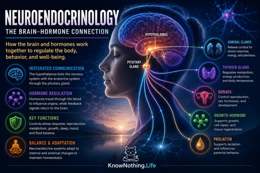
Neuroendocrinology is the study of communication between the nervous system and the endocrine system. It examines how neurons regulate hormone secretion, how circulating hormones alter brain activity, and how these interactions coordinate reproduction, stress, growth, metabolism, sleep, fluid balance, and behavior. The field challenges any simple division between the brain and the rest of the body. Neural activity can produce hormonal changes that affect distant organs, while hormones released by those organs return to the brain and modify motivation, mood, appetite, memory, and physiological priorities.
The hypothalamus occupies a central position in this communication network. This relatively small region of the forebrain receives information about light, temperature, energy availability, blood chemistry, emotional significance, and signals from internal organs. Specialized hypothalamic neurons translate those inputs into electrical and chemical messages that influence the pituitary gland. The pituitary then regulates endocrine organs such as the adrenal glands, thyroid, ovaries, and testes. Neuroendocrine control is therefore organized through interacting feedback loops rather than a one-way chain of commands.
The Hypothalamus and Pituitary Gland
The anterior pituitary is controlled partly by releasing and inhibiting hormones secreted from hypothalamic neurons into the hypophyseal portal circulation. These small blood vessels carry neurohormones directly from the hypothalamus to pituitary cells without first diluting them throughout the general circulation. Different hypothalamic signals stimulate or suppress the release of thyroid-stimulating hormone, adrenocorticotropic hormone, luteinizing hormone, follicle-stimulating hormone, growth hormone, and prolactin. The pituitary hormones then travel through the bloodstream to influence peripheral glands and tissues.
One of the discoveries that established this model was the identification of thyrotropin-releasing hormone, or TRH. In 1969, Roger Burgus, Thomas Dunn, Donald Desiderio, and Roger Guillemin reported evidence for the molecular structure of the hypothalamic thyrotropin-releasing factor. Burgus and colleagues subsequently characterized the purified ovine hormone in their 1970 study Characterization of Ovine Hypothalamic Hypophysiotropic TSH-Releasing Factor. The work demonstrated that a chemically identifiable peptide produced in the brain could control hormone secretion from the anterior pituitary, providing direct support for the central principle of neuroendocrinology.
The Neuroendocrine Stress Response
The hypothalamic-pituitary-adrenal axis, commonly called the HPA axis, coordinates one of the body’s principal responses to challenge. When neural systems interpret a situation as threatening or physiologically demanding, neurons in the hypothalamic paraventricular nucleus release corticotropin-releasing hormone, also known as corticotropin-releasing factor. This hormone stimulates pituitary corticotroph cells to release adrenocorticotropic hormone. ACTH then travels to the adrenal cortex, where it promotes the secretion of glucocorticoids, including cortisol in humans.
Wylie Vale, Joachim Spiess, Catherine Rivier, and Jean Rivier identified and characterized a 41-amino-acid ovine hypothalamic peptide that stimulated the secretion of ACTH and beta-endorphin in their landmark 1981 paper Characterization of a 41-Residue Ovine Hypothalamic Peptide That Stimulates Secretion of Corticotropin and β-Endorphin. Subsequent administration of synthetic CRF to human volunteers produced dose-dependent increases in ACTH and cortisol, confirming its functional role in the human stress axis. Glucocorticoids help mobilize energy and modify cardiovascular, immune, and cognitive activity, but they also feed back to the pituitary, hypothalamus, and other brain regions to restrain further activation.
Reproduction and Pulsatile Hormone Release
The hypothalamic-pituitary-gonadal axis regulates puberty, fertility, reproductive cycles, and sex-hormone production. Hypothalamic neurons release gonadotropin-releasing hormone, or GnRH, into the pituitary portal circulation. GnRH stimulates pituitary cells to secrete luteinizing hormone and follicle-stimulating hormone, which act on the ovaries or testes. These gonadotropins support the production of eggs and sperm while regulating the synthesis of estradiol, progesterone, and testosterone. The gonadal hormones then influence reproductive organs and return signals to the brain and pituitary.
A defining discovery was that the temporal pattern of GnRH is as important as its presence. In the 1978 study Hypophysial Responses to Continuous and Intermittent Delivery of Hypothalamic Gonadotropin-Releasing Hormone, Peter Belchetz, Tony Plant, Yasuhiro Nakai, Edward Keogh, and Ernst Knobil examined monkeys whose endogenous hypothalamic control had been interrupted. Intermittent GnRH restored sustained gonadotropin secretion, whereas continuous administration eventually suppressed pituitary responsiveness. The finding revealed that hormones carry information through timing and rhythm as well as concentration, and it later helped inform clinical uses of GnRH agonists and antagonists.
Oxytocin, Vasopressin, and the Posterior Pituitary
The posterior pituitary operates differently from the anterior pituitary. Oxytocin and vasopressin are synthesized by large hypothalamic neurons whose axons extend into the posterior pituitary. Electrical activity travels down these axons and triggers hormone release directly into the bloodstream. Vasopressin supports water conservation and cardiovascular regulation, while oxytocin is particularly important in uterine contractions and milk ejection. Both substances also act within the brain, where they can influence behavior and autonomic responses.
Vincent du Vigneaud and his colleagues determined the amino-acid sequence and proposed the chemical structure of oxytocin in 1953. They then produced a synthetic peptide with the biological activity of the natural hormone, an achievement reported in The Synthesis of an Octapeptide Amide with the Hormonal Activity of Oxytocin and expanded in The Synthesis of Oxytocin. This work was historically important not only for neuroendocrinology but also for peptide chemistry, because it showed that a pituitary hormone could be structurally defined and synthesized. Modern descriptions of oxytocin as a universal “love hormone,” however, oversimplify a molecule whose effects depend on neural context, receptor distribution, individual history, and the surrounding social environment.
Circadian Control of Hormone Secretion
Hormone secretion is organized in time. Cortisol tends to follow a daily pattern, melatonin rises in relation to darkness, growth hormone is strongly influenced by sleep, and reproductive hormones may be released in pulses or cycles. The principal mammalian circadian pacemaker is located in the suprachiasmatic nucleus, a small hypothalamic structure positioned above the optic chiasm. Light information reaching the SCN through retinal pathways helps synchronize internal physiology with the environmental day-night cycle.
Robert Moore and Victor Eichler demonstrated the SCN’s importance in their 1972 study Loss of a Circadian Adrenal Corticosterone Rhythm Following Suprachiasmatic Lesions in the Rat. Damage to this region abolished the normal daily corticosterone rhythm, showing that the SCN is required for temporal organization of an endocrine output. Circadian regulation is not merely a reaction to light at each moment. The SCN coordinates internal oscillations and communicates with neural and hormonal pathways that regulate sleep, temperature, feeding, autonomic activity, and endocrine secretion. Neuroendocrine health therefore depends not only on how much hormone is released but also on when it appears.
Metabolism and Signals from the Body
Neuroendocrine communication also allows the brain to monitor the body’s energy condition. Hormones from the pancreas, digestive tract, adrenal glands, and adipose tissue provide information about food intake, stored energy, glucose availability, and physiological demand. The hypothalamus integrates these signals with sensory experience, reward, stress, temperature, and learned expectations. Eating behavior is consequently regulated by a distributed system rather than a single hunger center.
The discovery of leptin transformed understanding of this process by showing that adipose tissue functions as an endocrine organ. Leptin circulates in relation to stored body fat and acts on neural systems involved in food intake, energy expenditure, and reproductive function. Louis Tartaglia and colleagues identified and cloned the leptin receptor, OB-R, in 1995, finding strong expression in regions including the hypothalamus. Research published the same year by Richard Maffei and colleagues showed that circulating leptin levels reflected body lipid content, while animals fed a high-fat diet developed reduced responsiveness despite elevated hormone levels. These findings helped establish the concept that endocrine disorders can involve resistance to a signal rather than simple hormone deficiency.
Feedback, Plasticity, and Clinical Importance
Neuroendocrine systems depend on feedback. Thyroid hormones inhibit further activation of the hypothalamic-pituitary-thyroid axis, glucocorticoids restrain parts of the stress response, and gonadal steroids regulate GnRH and gonadotropin secretion. Feedback can be negative, reducing further hormone release, or positive in specialized circumstances, such as the reproductive signals contributing to the preovulatory luteinizing-hormone surge. Receptors, binding proteins, hormone metabolism, and neural context determine how strongly a signal affects a particular cell.
Disorders can arise at every level of these systems. A problem may originate in a hypothalamic neuron, the pituitary, a peripheral gland, a receptor, or a feedback pathway. Neuroendocrinology therefore contributes to the understanding and treatment of infertility, thyroid disease, pituitary tumors, stress-related disorders, growth abnormalities, metabolic disease, and disturbances of fluid balance. Its larger importance lies in revealing that thought, environment, physiology, and behavior are biologically connected. The brain regulates the body through hormones, while hormones continually reshape the brain’s priorities and responses.
Why Neuroendocrinology Matters
Neuroendocrinology provides a framework for understanding how the organism acts as a coordinated whole. A stressful event can change cortisol secretion; light can alter circadian hormone rhythms; nutritional status can influence reproduction; and reproductive hormones can modify neural activity and behavior. None of these processes belongs entirely to either neuroscience or endocrinology. They emerge from communication across both systems.
The field’s major discoveries have shown that endocrine messages possess structure, timing, location, and context. Hormones may be secreted in pulses, released according to circadian schedules, transported through specialized blood vessels, or delivered directly from neural terminals. Their effects are shaped by feedback and by the state of the receiving tissue. Neuroendocrinology ultimately explains how the brain translates experience into bodily regulation—and how the body, through its hormones, changes the way the brain perceives and responds to the world.


