
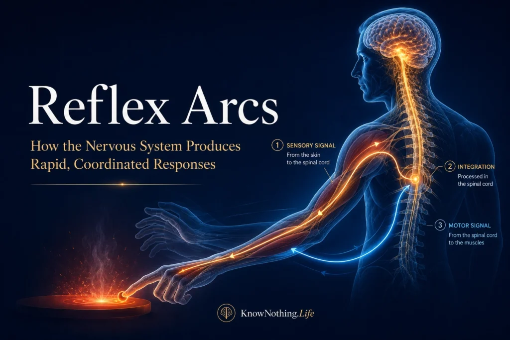
A reflex arc is a neural pathway that transforms sensory information into a rapid, involuntary response. The familiar example is the knee-jerk reflex, but reflex circuits participate in far more than dramatic reactions to a tendon tap. They help maintain posture, stabilize joints, regulate muscle tension, protect the body from injury, control pupil size, adjust blood pressure, and coordinate movements during walking. A reflex does not necessarily occur without involvement from the brain. Some reflexes are integrated primarily within the spinal cord, while others depend on the brainstem or autonomic centers. Even spinal reflexes remain subject to signals descending from the brain, allowing attention, emotion, movement, and experience to change their strength.
The modern understanding of reflexes owes much to Charles Scott Sherrington, whose experiments showed that the spinal cord is not merely a bundle of conducting fibers. In papers such as “Observations on Some Spinal Reflexes and the Interconnection of Spinal Segments” and “Flexion-Reflex of the Limb, Crossed Extension-Reflex, and Reflex Stepping and Standing,” Sherrington demonstrated that sensory inputs could activate coordinated patterns of excitation and inhibition across multiple spinal segments. His 1906 book, The Integrative Action of the Nervous System, presented the nervous system as an organized network in which reflexes cooperate, compete, and combine to produce adaptive behavior.
Components of a Reflex Arc
The standard reflex arc contains five functional components: a sensory receptor, an afferent neuron, an integration center, an efferent neuron, and an effector. A receptor first detects a meaningful change, such as muscle stretch, skin pressure, heat, tissue damage, light, or altered blood pressure. The receptor converts that event into electrical activity that travels along an afferent nerve fiber toward the spinal cord or brainstem. Inside the central nervous system, the afferent signal communicates either directly with a motor neuron or indirectly through one or more interneurons. An efferent pathway then carries the resulting command to a muscle, gland, blood vessel, or organ.
This textbook sequence is useful, but real reflex pathways are rarely isolated chains. One sensory fiber may branch and influence motor neurons, inhibitory interneurons, ascending sensory pathways, and neighboring spinal segments at the same time. The response may therefore include contraction of one muscle, relaxation of its antagonist, stabilization of another joint, and transmission of information to the brain. Sherrington called motor neurons the “final common path” because commands from reflex circuits, voluntary motor systems, and other neural sources must converge on the neurons that activate skeletal muscle. His work on reflex inhibition emphasized that coordinated movement requires the nervous system to suppress inappropriate activity as carefully as it generates excitation.
The Stretch Reflex and Muscle Spindles
The stretch reflex helps muscles resist sudden lengthening and contributes to posture, balance, and joint stability. Its receptors are muscle spindles, specialized sensory structures located within skeletal muscles. When a muscle lengthens, spindle afferents increase their firing and transmit signals into the spinal cord. The fastest pathway allows group Ia sensory fibers to excite alpha motor neurons supplying the same muscle. The activated motor neurons cause the stretched muscle to contract, opposing the original change in length. This direct connection is why the central excitatory component of the stretch reflex is described as monosynaptic.
The complete response is more complex than a single synapse. Ia afferents also activate inhibitory interneurons that reduce activity in motor neurons supplying the opposing muscle, a process known as reciprocal inhibition. When the quadriceps contracts during the patellar reflex, inhibition of the hamstrings allows the lower leg to extend with less opposition. Muscle-spindle sensitivity is also adjusted by gamma motor neurons, while signals from the skin, joints, other muscles, and brain modify spinal excitability. Studies of mechanically induced responses have identified both short-latency components associated with the conventional stretch pathway and longer-latency responses involving additional spinal or supraspinal processing.
Clinicians and researchers can examine similar circuitry through the Hoffmann reflex, or H-reflex. Instead of stretching a muscle spindle with a tendon tap, electrical stimulation activates sensory fibers within a mixed peripheral nerve. The resulting signal travels to the spinal cord, excites motor neurons, and produces a measurable muscle response. Because the H-reflex bypasses the mechanical behavior of the spindle and tendon, it is not identical to a tendon jerk. Nevertheless, it provides a valuable indirect measure of spinal reflex excitability, presynaptic inhibition, motor-neuron responsiveness, and descending control.
Withdrawal and Crossed-Extension Reflexes
The withdrawal reflex protects the body from potentially damaging stimulation. Nociceptive sensory fibers activated by excessive heat, sharp pressure, or tissue injury enter the spinal cord and engage networks of interneurons. These circuits activate muscles capable of moving the affected body part away from the stimulus while inhibiting muscles that would oppose withdrawal. Because the appropriate response depends on where the stimulus occurs, withdrawal is not simply a universal contraction of every flexor muscle. Stimulation on one part of the foot may require a different combination of hip, knee, and ankle movements than stimulation at another location.
Research led by Jens Schouenborg helped establish that withdrawal circuits have a modular organization. Each module links an area of the skin with muscles capable of moving that area away from danger efficiently. This arrangement creates a functional spinal representation of the body based on protective action rather than sensation alone. Human experiments further show that the nervous system integrates nociceptive information across space and time before determining the magnitude of the response. A stronger or more widespread stimulus can recruit a larger withdrawal response, while descending inhibitory systems can reduce spinal nociceptive transmission.
When withdrawal threatens balance, the opposite limb may produce a crossed-extension response. If one leg flexes away from a painful stimulus, spinal pathways can increase extensor activity in the other leg so it can support the body. Sherrington’s experiments demonstrated that flexion, crossed extension, stepping, and standing emerge from coordinated combinations of excitation and inhibition rather than independent reflex switches. The exact pattern varies with posture, sensory context, and the state of the nervous system, showing that even protective responses are integrated with the larger task of maintaining stability.
Reflexes During Posture and Movement
Reflexes are sometimes presented as reactions that interrupt voluntary movement, but they are also built into movement itself. While standing, continuous sensory information from muscle spindles, tendons, skin, joints, the vestibular system, and vision helps the nervous system detect sway and adjust muscle activity. During walking, the effect of a sensory stimulus changes according to the phase of the step cycle. A cutaneous input that assists foot clearance during the swing phase may produce a different response when the foot is supporting body weight. This phase-dependent modulation prevents a fixed reflex from disrupting locomotion.
Experiments on cutaneous reflexes have shown that their strength and pattern change with walking speed, supporting the view that spinal circuits adapt sensory feedback to current movement demands. Hip position can also change the excitability of reflex pathways affecting muscles around the ankle, demonstrating communication among different joints and spinal segments. Reflexes therefore operate within a task-dependent control system. The same sensory input can generate different outputs depending on whether a person is standing, walking, running, reaching, or recovering from a disturbance.
Reflex Modulation, Learning, and Plasticity
Reflex strength is not permanently fixed. Attention, anticipation, voluntary contraction, fatigue, pain, posture, and descending signals from the brain can alter the probability that motor neurons will respond to sensory input. The Jendrassik maneuver illustrates this modulation in clinical practice. A person interlocks the fingers and pulls while a clinician tests a leg reflex, often making a weak tendon response easier to observe. Studies of the maneuver suggest that it changes spinal excitability and presynaptic control rather than merely distracting the person from the tendon tap.
Repeated activity can also change reflex pathways. H-reflex responses may decrease with repeated stimulation, demonstrating habituation, while training can modify reflex excitability according to a motor task’s requirements. In a study of people with incomplete spinal cord injuries, Aiko Thompson and colleagues used operant conditioning to reduce an abnormally large soleus H-reflex. Participants who successfully changed the reflex also showed improvements in walking speed and symmetry. The results indicate that a spinal reflex can be shaped through learning and that modifying one pathway may encourage broader improvements across the motor system.
Autonomic and Cranial Reflex Arcs
Not all reflex arcs end in skeletal muscle. Autonomic reflexes regulate internal organs, glands, smooth muscle, and circulation. In the arterial baroreflex, pressure-sensitive receptors in major arteries detect changes in vessel stretch. Sensory signals reach cardiovascular centers in the brainstem, which adjust sympathetic and parasympathetic activity to the heart and blood vessels. This pathway helps stabilize circulation when a person stands, exercises, sleeps, or experiences emotional stress. Like spinal reflexes, the baroreflex is continuously modified according to behavioral context rather than operating around one unchanging set point.
The pupillary light reflex is another brainstem-mediated pathway. Light activates retinal cells whose signals reach pretectal regions of the midbrain. Bilateral connections then influence parasympathetic neurons associated with the oculomotor nerves, producing constriction in both the illuminated pupil and the opposite pupil. Because the pathway contains distinct afferent and efferent components, abnormal pupil responses can help clinicians localize damage affecting the retina, optic nerve, midbrain, oculomotor nerve, or peripheral parasympathetic fibers. Experimental tracing and stimulation studies have helped define the pathway connecting visual input with the ciliary ganglion and iris sphincter.
Clinical Importance of Reflex Testing
Reflex examination gives clinicians a rapid way to assess the integrity of sensory nerves, spinal segments, motor neurons, neuromuscular junctions, muscles, and descending pathways. A diminished tendon reflex may result from injury to the sensory or motor portion of the arc, peripheral neuropathy, nerve-root compression, or motor-neuron dysfunction. Exaggerated reflexes may appear when damage to the brain or spinal cord reduces descending inhibition. Clonus, spasticity, and expansion of reflex receptive fields can reflect abnormal excitability within spinal networks rather than simply stronger muscles.
A reflex result must always be interpreted in context. Reflex strength varies among healthy people and can change with age, anxiety, reinforcement maneuvers, medication, body position, and testing technique. Comparisons between the two sides of the body and relationships among several reflexes are often more informative than one response considered alone. Reflex arcs are therefore not primitive shortcuts operating separately from thought and movement. They are adaptable sensorimotor and autonomic networks through which the nervous system protects the body, preserves stability, regulates internal conditions, and continually adjusts behavior to a changing world.


