
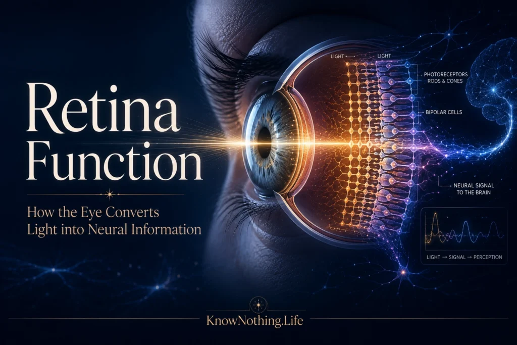
The retina is a thin, layered sheet of neural tissue lining the back of the eye. Although it is often compared to the sensor in a camera, it does much more than receive an image. The retina converts light into electrical activity, adjusts its sensitivity to changing illumination, separates increases from decreases in brightness, compares neighboring regions of visual space, and extracts information about color, contrast, movement, and timing. By the time visual signals leave the eye through the optic nerve, they have already undergone substantial processing. The brain therefore does not receive a raw photograph of the environment. It receives multiple streams of encoded information produced by specialized retinal circuits.
The retina develops as an extension of the central nervous system and contains several major classes of neurons: photoreceptors, horizontal cells, bipolar cells, amacrine cells, and retinal ganglion cells. These cells are arranged in layers connected by two major synaptic regions. Light passes through much of the neural tissue before reaching the photoreceptors near the retinal pigment epithelium. Neural signals then travel in the opposite direction, from photoreceptors through interneurons to ganglion cells. The ganglion-cell axons gather at the optic disc, form the optic nerve, and carry retinal output toward the thalamus, superior colliculus, hypothalamus, and other brain structures.
Rods, Cones, and Phototransduction
Vision begins in rods and cones, the retina’s primary photoreceptors. Rods are highly sensitive and dominate vision under dim conditions, but they provide relatively limited spatial and color information. Cones function most effectively under brighter illumination and support detailed vision, rapid responses, and color discrimination. Humans possess three major cone classes with different spectral sensitivities. Rather than identifying colors individually, these cones provide signals that are compared by later retinal circuits. Their distribution is uneven: cones are densely concentrated in the central retina, especially the fovea, while rods are more numerous outside the foveal center.
Phototransduction is the process through which absorbed light changes a photoreceptor’s electrical state. In darkness, cyclic guanosine monophosphate helps keep ion channels open, producing a steady inward current and continuous glutamate release. Light activates an opsin-containing visual pigment and initiates a biochemical cascade that lowers cyclic GMP, closes channels, and hyperpolarizes the cell. Photoreceptors therefore reduce their neurotransmitter release when illuminated. Dennis Baylor, Trevor Lamb, and King-Wai Yau demonstrated the extraordinary sensitivity of this system in their 1979 study “Responses of Retinal Rods to Single Photons,” showing that individual rods can produce measurable responses to single absorbed photons.
From Photoreceptors to ON and OFF Pathways
Photoreceptors communicate with bipolar and horizontal cells in the outer retina. Horizontal cells link signals across neighboring photoreceptors and contribute to spatial comparison, adaptation, and receptive-field surrounds. Bipolar cells carry information vertically toward the inner retina, but they do not all respond to light in the same way. ON bipolar cells become more active when illumination increases, while OFF bipolar cells become more active when illumination decreases. This division allows the retina to represent brightening and darkening through parallel channels rather than relying on a single signal that merely rises or falls.
The ON pathway depends strongly on the metabotropic glutamate receptor mGluR6. In darkness, glutamate released by photoreceptors activates this receptor and suppresses ON bipolar-cell activity. When light reduces glutamate release, the suppression is relieved and the ON bipolar cell depolarizes. OFF bipolar cells use different glutamate receptors and preserve the photoreceptor signal’s sign, responding when glutamate release increases as light decreases. Experiments disrupting the mGluR6 gene produced a selective loss of normal ON responses while leaving OFF transmission comparatively intact, demonstrating that the two channels depend on distinct synaptic mechanisms.
Contrast and Center-Surround Receptive Fields
One of the retina’s central functions is to emphasize contrast. A ganglion cell does not respond equally to every light falling within a broad area. Instead, it has a receptive field: a region of visual space in which stimulation changes its activity. Many retinal ganglion cells possess center-surround receptive fields. An ON-center cell is excited when light appears in the center and inhibited when light illuminates the surrounding region. An OFF-center cell displays the reverse pattern. Uniform illumination across both center and surround may create a weaker response because the opposing influences partially cancel one another.
Stephen Kuffler established the importance of this organization in his landmark 1953 paper “Discharge Patterns and Functional Organization of Mammalian Retina.” Recording from mammalian retinal ganglion cells, Kuffler described ON-center, OFF-center, and ON-OFF response patterns and demonstrated that retinal output depends on the spatial arrangement of illumination. Center-surround organization enables the retina to emphasize borders, local differences, shadows, and changes rather than simply reporting absolute light intensity. This efficient coding helps preserve visually useful information while reducing redundant signals from uniform surfaces.
Amacrine Cells, Timing, and Motion
Amacrine cells form an exceptionally diverse group of retinal interneurons. They influence communication between bipolar cells and ganglion cells and contribute to temporal sensitivity, contrast adaptation, motion detection, and the separation of rod and cone pathways. Some amacrine cells respond briefly when a stimulus appears or disappears, helping the retina distinguish rapid change from sustained illumination. Others connect signals across larger retinal distances or release inhibitory neurotransmitters that shape when and where ganglion cells fire. The retina consequently analyzes the timing of visual events as well as their spatial structure.
Starburst amacrine cells are especially important in retinal direction selectivity. Their radially arranged dendrites release acetylcholine and the inhibitory transmitter GABA and connect asymmetrically with direction-selective ganglion cells. These circuits help certain ganglion cells respond strongly when an image moves in one direction but weakly when it moves in the opposite direction. In a 2001 experiment, Keisuke Yoshida and colleagues selectively eliminated starburst amacrine cells and found that normal direction-selective ganglion-cell responses were severely disrupted. The study demonstrated that meaningful motion analysis begins within the retina rather than occurring exclusively in the visual cortex.
Retinal Ganglion Cells and Parallel Visual Messages
Retinal ganglion cells are the retina’s output neurons. They receive processed input from bipolar and amacrine cells and convert it into action potentials that travel along the optic nerve. Rather than forming one uniform population, ganglion cells belong to numerous anatomical and functional types. Each type samples the visual scene differently and sends a distinctive message to particular brain targets. Some emphasize fine detail, some respond strongly to movement or rapid contrast changes, and others contribute to color, orientation, looming detection, or the distinction between object motion and motion affecting the entire visual field.
In primates, midget and parasol ganglion cells provide major output pathways. Midget cells generally have relatively small receptive fields, especially near the fovea, and contribute strongly to high-resolution spatial vision and red-green color comparisons. Parasol cells have larger receptive fields and respond effectively to luminance contrast, movement, and rapid temporal changes. Small bistratified ganglion cells contribute to a blue-ON color-opponent pathway by comparing short-wavelength cone signals with combinations of medium- and long-wavelength cone activity. Dennis Dacey and Barry Lee linked this pathway to a morphologically distinct ganglion-cell class, illustrating how retinal anatomy and visual function are organized into parallel channels.
Color Vision and Retinal Comparison
Color vision depends on comparisons among cone signals rather than on isolated responses from individual photoreceptors. Long-, medium-, and short-wavelength-sensitive cones have overlapping response ranges, meaning that a cone’s activity alone cannot identify a wavelength unambiguously. Retinal circuits compare these signals through opponent pathways. Some neurons contrast long- and medium-wavelength cone activity, supporting red-green discrimination, while other pathways compare short-wavelength input with combined long- and medium-wavelength signals, contributing to blue-yellow discrimination.
Color opponency is distributed across several retinal cell types and cannot be reduced to one simple circuit. The small bistratified pathway provides a well-characterized example in which short-wavelength cone input produces an ON response while opposing cone signals produce an OFF response. Midget pathways also contribute to the high-resolution chromatic information associated with primate vision, although their exact mechanisms can vary with retinal location and patterns of cone input. The retina thus converts overlapping photoreceptor responses into relational signals that the brain can use to distinguish surfaces under changing conditions.
Light Adaptation and Vision Across Changing Conditions
The visual environment can vary from near darkness to intense daylight, producing changes in illumination that exceed the response range of any single neuron. The retina compensates through adaptation at several levels. Photoreceptors adjust the gain and timing of their responses, while horizontal, bipolar, amacrine, and ganglion-cell circuits alter their sensitivity to local brightness and contrast. Rod pathways dominate under very dim conditions, mixed rod-and-cone activity supports intermediate illumination, and cone pathways become increasingly important in daylight. Adaptation prevents retinal responses from becoming permanently saturated and helps preserve sensitivity to meaningful changes.
Retinal adaptation is also local and feature dependent. Ganglion cells can adjust their responses to variations in contrast, motion, color statistics, and the recent history of stimulation. Studies using natural scenes show that retinal activity cannot always be predicted perfectly from responses to simple spots or flashes. Eye movements, textures, correlations, and changing visual contexts influence how retinal circuits encode information. Modern models trained on naturalistic stimuli have reproduced phenomena involving adaptation, motion sensitivity, and context, reinforcing the view that the retina is tuned to the statistical structure of the environments in which vision normally operates.
Vision Beyond Conscious Image Perception
Not all retinal functions contribute directly to conscious visual images. A small population of retinal ganglion cells contains melanopsin and is intrinsically sensitive to light. These intrinsically photosensitive retinal ganglion cells also receive signals from rods and cones, but their own photopigment allows them to respond directly, especially to sustained illumination. Their projections influence the biological clock, pupil constriction, sleep regulation, hormonal rhythms, and other physiological responses to environmental light.
Samer Hattar and colleagues described the architecture and intrinsic photosensitivity of melanopsin-containing ganglion cells in 2002, showing that they project to the suprachiasmatic nucleus, the brain’s central circadian clock. Studies of mice lacking functional melanopsin later found weakened pupillary responses at high light intensities, confirming that melanopsin contributes substantially to non-image-forming responses. These discoveries expanded the meaning of retinal function: the retina does not merely help an organism see objects but also informs the body about the timing and intensity of environmental light.
The Retina as an Active Visual Computer
The retina should not be understood as passive film placed at the back of the eye. It is an active neural processor that compresses, separates, and reorganizes visual information before that information reaches the brain. Photoreceptors convert photons into graded electrical signals; horizontal and bipolar cells establish spatial and ON-OFF comparisons; amacrine cells shape timing and motion responses; and ganglion cells divide the results into parallel output channels. Every stage contributes to deciding which aspects of the visual world will be transmitted.
Modern retinal research combines electrophysiology, microscopy, genetics, computational modeling, and large-scale recordings from many cells at once. These methods are identifying increasingly specific retinal cell types and revealing how their responses change under natural viewing conditions. The findings guide research into retinal degeneration, artificial vision, gene therapy, prosthetic devices, and strategies for restoring communication between the eye and brain. Yet the retina’s basic achievement remains remarkable: from the absorption of individual photons, a compact neural network creates organized signals capable of supporting sight, movement, biological timing, and the brain’s continuing construction of the visible world.


