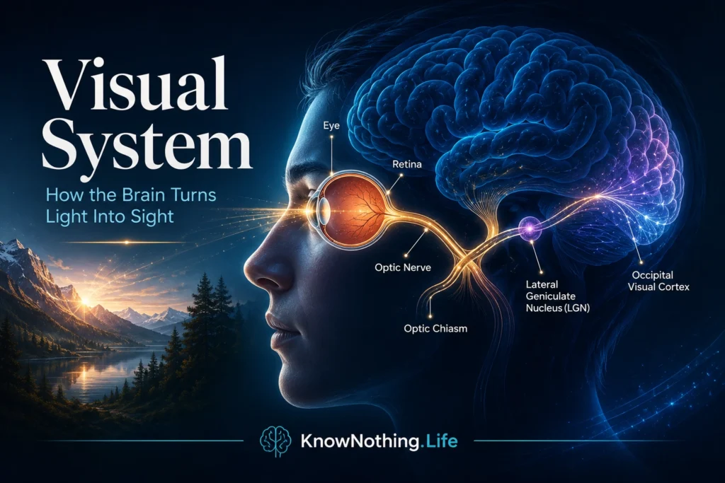
The visual system is the biological network that allows the brain to detect light, process visual information, and create the experience of seeing. It includes the eyes, retina, optic nerves, optic chiasm, optic tracts, lateral geniculate nucleus of the thalamus, optic radiations, primary visual cortex, and higher visual pathways that extend into the temporal and parietal lobes. A medical overview of the visual pathway describes it as a continuous series of structures carrying visual information from the retina through the optic nerve, optic chiasm, optic tract, lateral geniculate nucleus, optic radiations, and finally the occipital lobe.
Vision may feel immediate, but it is not a simple camera-like recording of the world. Light enters the eye, but sight is built by the nervous system. The brain must detect contrast, color, motion, depth, edges, orientation, faces, objects, symbols, and spatial relationships. It must also connect visual information with memory, attention, emotion, language, and movement. The visual system therefore does more than show us what is “out there.” It transforms light into perception, perception into meaning, and meaning into action.
The Eye and Retina
The visual process begins when light enters the eye through the cornea and pupil, passes through the lens, and is focused onto the retina at the back of the eye. The retina is not just a passive screen. It is neural tissue containing photoreceptors and several layers of processing cells. Rods are especially sensitive to low light and support night vision, while cones support color and high-acuity vision. Retinal circuits begin processing contrast, edges, brightness, motion, and spatial information before signals ever leave the eye.
Photoreceptors communicate with bipolar cells, horizontal cells, amacrine cells, and retinal ganglion cells. The axons of retinal ganglion cells form the optic nerve, which carries visual signals toward the brain. This means the eye is already part of the nervous system’s processing architecture. Seeing does not begin in the cortex alone. The retina performs early analysis, compressing and organizing visual information so the brain receives structured signals rather than raw light. The visual system is therefore a distributed pathway, not a single organ.
The Visual Pathway to the Brain
After leaving the retina, visual information travels through the optic nerves toward the optic chiasm. At the chiasm, fibers from the nasal halves of the retinas cross to the opposite side, while fibers from the temporal halves remain on the same side. This arrangement allows visual information from the left visual field to be processed mainly by the right hemisphere and information from the right visual field to be processed mainly by the left hemisphere. From there, signals travel through the optic tracts toward the lateral geniculate nucleus, or LGN, of the thalamus.
The LGN is often described as a relay station, but it is more than a passive stop along the way. A StatPearls review describes the LGN as a sensory projection nucleus of the thalamus that plays an essential role in visual processing and has broad connectivity with visual cortical regions. From the LGN, signals travel through the optic radiations to the primary visual cortex in the occipital lobe. This pathway is organized retinotopically, meaning neighboring points in the visual field are represented in neighboring areas of the brain. NCBI’s neuroscience text notes that the primary pathway from retina to LGN to primary visual cortex is the most thoroughly studied component of the visual system.
The Visual Cortex and Feature Processing
The primary visual cortex, also called V1 or striate cortex, is located in the occipital lobe. It is the first major cortical region devoted to visual processing. StatPearls describes the visual cortex as the primary cortical region that receives, integrates, and processes visual information relayed from the retinas, and notes that visual cortex is often divided into areas such as V1 through V5 based on structure and function. In V1, the brain begins a more detailed analysis of visual features such as edges, contrast, orientation, spatial frequency, and location.
The classic work of David Hubel and Torsten Wiesel transformed modern neuroscience by showing that neurons in the visual cortex respond selectively to features such as line orientation and that visual cortex is organized into functional columns. Their research helped establish that the cortex actively constructs visual perception by extracting features from incoming information. Eric Kandel later wrote that Hubel and Wiesel not only opened the study of primary visual cortex but laid the foundation for research across sensory systems. Their discoveries showed that seeing is an organized neural achievement, not the simple arrival of an image inside the brain.
Color, Motion, Depth, and Visual Specialization
After early processing in V1, visual information spreads into surrounding extrastriate regions. Different areas contribute to different visual features. Some regions are especially important for color, others for motion, depth, shape, texture, or object boundaries. This division of labor allows the visual system to break the world into components and then integrate those components into a coherent scene. The world appears unified, but the brain builds that unity from many specialized processes working together.
Motion processing is especially important because living organisms must detect movement quickly. A moving object may be prey, predator, vehicle, person, gesture, or threat. Depth perception allows the brain to estimate distance, guide reaching, avoid obstacles, and navigate space. Color helps identify surfaces, objects, emotional cues, food, warning signals, and environmental conditions. These features are not decorative additions to sight. They are functional tools the brain uses to interpret the world and guide behavior.
The Dorsal and Ventral Visual Streams
One of the most influential ideas in visual neuroscience is the two-stream model. According to Melvyn Goodale and David Milner’s 1992 proposal, the ventral stream projects from visual cortex toward the temporal lobe and plays a major role in identifying objects, while the dorsal stream projects toward the posterior parietal region and supports sensorimotor transformations for visually guided action. In simplified terms, the ventral stream helps answer “What is it?” while the dorsal stream helps answer “Where is it, and how should I act toward it?”
This distinction helps explain why visual perception and visual action can sometimes dissociate. A person may struggle to consciously identify an object yet still shape the hand appropriately when reaching for it, or may recognize an object but have difficulty using visual information to guide movement. The ventral stream contributes to object recognition, faces, forms, and meaning. The dorsal stream contributes to spatial awareness, reaching, grasping, eye movements, and navigation. Vision is therefore not one single ability. It is a family of linked processes that help the brain recognize the world and act within it.
Visual Attention and Perception
The visual system receives far more information than the brain can consciously process in detail. Attention helps select what becomes clear, relevant, and actionable. When a person looks for a friend in a crowd, reads a sentence, drives through traffic, or notices a sudden movement, visual attention is shaping perception. The thalamus, visual cortex, parietal lobe, frontal eye fields, and other networks all help determine where the eyes move and what the mind notices.
This is why sight and attention cannot be completely separated. The eyes may receive information from a scene, but awareness depends on selection. People can miss obvious visual events when attention is elsewhere, and the same visual scene can be interpreted differently depending on expectation, task, emotion, and memory. The visual system does not merely detect light. It organizes visual experience according to relevance, context, and action.
Clinical Importance of the Visual System
Damage anywhere along the visual pathway can produce different symptoms. Damage to the retina or optic nerve may impair vision in one eye. Damage at the optic chiasm can produce characteristic field losses. Damage to optic radiations or the occipital cortex can cause visual-field deficits even when the eyes themselves are healthy. Damage to higher visual areas can affect object recognition, face recognition, motion perception, spatial awareness, reading, or visually guided action.
These clinical patterns reveal the visual system’s organization. Vision is not located in one place. It depends on pathways, relays, cortical maps, and distributed streams. A person may have normal eyes but impaired cortical vision. Another may see objects but fail to recognize them. Another may recognize objects but struggle to locate or reach for them accurately. These conditions show that vision includes detection, perception, recognition, attention, meaning, and action.
Why the Visual System Matters
The visual system matters because vision is one of the main ways humans understand and act in the world. It allows people to read, recognize faces, navigate streets, judge distance, appreciate art, detect danger, interpret expression, and coordinate movement. But its deeper importance is that it reveals how the brain creates experience. Light itself is not sight. Sight emerges when neural systems transform light into patterns, patterns into objects, objects into meanings, and meanings into behavior.
The visual system also teaches one of neuroscience’s central lessons: perception is active. The brain does not simply receive the world as a finished picture. It builds a usable world through receptors, pathways, thalamic relays, cortical maps, attention, memory, and action systems. To understand the visual system is to understand how biology turns energy into awareness and how the brain transforms light into the visible world we live in.