
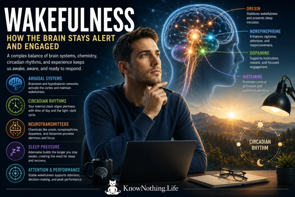
Wakefulness is the brain state in which a person is capable of responding to the environment, directing attention, producing voluntary behavior, and maintaining an organized relationship with the surrounding world. It is often treated as the opposite of sleep, but wakefulness is not a single uniform condition. A person can be intensely alert, quietly attentive, distracted, drowsy, or behaviorally awake while experiencing brief lapses in information processing. Wakefulness therefore varies in stability and quality even when the eyes remain open and the body continues to move.
Wakefulness is also distinct from consciousness. Wakefulness describes the level of physiological arousal, whereas consciousness refers more broadly to subjective experience and awareness. Most conscious experiences occur during wakefulness, but vivid dreaming demonstrates that consciousness can persist during sleep. Conversely, a person with unresponsive wakefulness syndrome may open the eyes and display sleep–wake cycles without producing reliable behavioral evidence of awareness. Neuroscience must therefore explain both how the brain awakens and how it organizes meaningful conscious experience once wakefulness has been established.
The Discovery of the Brain’s Arousal Systems
An important turning point came in 1949, when Giuseppe Moruzzi and Horace Magoun published “Brain Stem Reticular Formation and Activation of the EEG.” By electrically stimulating regions of the brainstem reticular formation, they changed the cerebral cortex from the slower, synchronized electrical activity associated with sleep or reduced arousal to the faster, lower-amplitude pattern associated with wakefulness. Their work helped establish the concept of an ascending reticular activating system: a set of brainstem pathways capable of influencing the thalamus and widespread areas of the cerebral cortex.
Wakefulness is now understood to depend on several interacting arousal systems rather than one anatomical switch. Neurons in the brainstem, hypothalamus, basal forebrain, and thalamus release neurotransmitters that alter cortical excitability, sensory processing, motivation, and attention. These populations include noradrenergic neurons in the locus coeruleus, serotonergic neurons in the raphe nuclei, histaminergic neurons in the tuberomammillary nucleus, dopaminergic populations in the midbrain, cholinergic neurons in the brainstem and basal forebrain, and orexin-producing neurons in the lateral hypothalamus. Their projections overlap, but each system contributes differently to alertness, behavioral flexibility, attention, movement, and the stability of the waking state.
Orexin and the Stability of Wakefulness
Orexin, also called hypocretin, is one of the most important regulators of stable wakefulness. Takeshi Sakurai and colleagues identified orexin-A and orexin-B in 1998 and located the neurons producing them within the lateral and posterior hypothalamus. Although the number of orexin neurons is relatively small, their axons reach many wake-promoting regions, including the locus coeruleus, tuberomammillary nucleus, raphe nuclei, basal forebrain, and cerebral cortex. Orexin signaling appears especially important when an organism must maintain wakefulness because of motivation, hunger, danger, emotional stimulation, or environmental opportunity.
Antoine Adamantidis and colleagues provided causal evidence in their 2007 study “Neural Substrates of Awakening Probed with Optogenetic Control of Hypocretin Neurons.” Selectively stimulating orexin neurons in sleeping mice increased the probability that the animals would transition into wakefulness from either non-rapid eye movement or rapid eye movement sleep. Orexin does not merely produce generalized excitation; it helps stabilize boundaries between behavioral states. Human and animal narcolepsy research reinforces this conclusion. Studies of people with narcolepsy and cataplexy have found a severe loss of hypocretin signaling, while examinations of human brain tissue identified a generalized absence of hypocretin peptides in affected patients.
Neurotransmitters and the Waking Brain
The locus coeruleus supplies much of the brain’s norepinephrine and is strongly associated with arousal, vigilance, and responses to important events. Matthew Carter and colleagues used optogenetic methods to show that activating locus coeruleus neurons rapidly increased the probability of awakening, while inhibiting them reduced wakefulness. The effects depended on the frequency of stimulation, demonstrating that norepinephrine-producing neurons can causally regulate arousal rather than merely becoming active after awakening has already occurred.
Histamine, acetylcholine, serotonin, and dopamine make additional contributions. Histaminergic neurons in the hypothalamic tuberomammillary nucleus are most active during wakefulness, and experimental silencing of these cells can promote slow-wave sleep under certain conditions. Acetylcholine supports cortical activation, attention, learning, and sensory responsiveness, while dopamine contributes to motivation and the willingness to sustain effort. These chemical systems do not operate independently. Orexin neurons activate several of them, inhibitory sleep-promoting circuits suppress them, and feedback among arousal centers prevents either sleep or wake activity from becoming uncontrolled. Experimental work has even shown that wake-active histamine neurons release both excitatory histamine and inhibitory GABA-related signals, illustrating how arousal systems contain internal braking mechanisms.
Circadian Timing and the Drive to Stay Awake
The likelihood of remaining awake depends partly on the circadian timing system. The suprachiasmatic nucleus of the hypothalamus receives information about environmental light and coordinates daily rhythms in melatonin, body temperature, hormone release, metabolism, and behavior. During the biological day, circadian signals provide an increasing drive for wakefulness that counteracts the sleep pressure accumulating with every hour spent awake. This alerting influence often becomes especially strong in the hours before habitual bedtime, creating what researchers call the wake-maintenance zone.
Experiments that separated the imposed sleep schedule from the internal circadian rhythm showed that alertness and cognitive performance are controlled by both time awake and biological time. Derk-Jan Dijk, Charles Czeisler, and their colleagues found that performance declines as prior wakefulness increases, but the circadian system can temporarily oppose this deterioration during the habitual waking day. Alertness and performance are generally strongest near the circadian peak in core body temperature and weakest near the biological night, when melatonin is elevated and temperature approaches its minimum. The same amount of prior wakefulness can therefore produce very different levels of functioning depending on circadian phase.
Sleep Pressure, Adenosine, and the Need for Recovery
Alexander Borbély’s influential two-process model describes sleep and wakefulness as the interaction of a homeostatic process and a circadian process. The homeostatic component, commonly called Process S, represents sleep pressure that builds during wakefulness and declines during sleep. The circadian component, Process C, changes according to biological time and alternately promotes sleep or wakefulness. Their interaction explains why a well-rested person can remain alert for much of the day, why sleepiness rises late at night, and why a person kept awake overnight may temporarily feel more alert again when the morning circadian signal increases.
Adenosine is one biological signal associated with accumulating sleep pressure. It is produced in relation to cellular energy use and influences neurons through several receptor types. Tarja Porkka-Heiskanen and colleagues found that extracellular adenosine increased in the basal forebrain during prolonged wakefulness and declined during recovery sleep. Adenosine can inhibit wake-promoting neurons, reducing cortical activation and increasing the likelihood of sleep. Caffeine promotes alertness largely by blocking adenosine receptors, but it does not eliminate the underlying need for sleep. It temporarily reduces the expression of sleep pressure while the physiological consequences of extended wakefulness continue to accumulate.
Attention, Performance, and Waking Lapses
Stable wakefulness is necessary for sustained attention, but people do not always recognize when their performance is deteriorating. The psychomotor vigilance test has been widely used to measure reaction speed and attentional lapses during sleep deprivation. Research shows that extended wakefulness slows responses, increases errors, and creates occasional long lapses in which a person fails to respond promptly. These failures are especially dangerous during driving, medical work, industrial operations, and other activities requiring continuous monitoring.
Chronic partial sleep loss can be as disruptive as a single night without sleep. Hans Van Dongen, David Dinges, and colleagues restricted healthy adults to four, six, or eight hours in bed for fourteen nights. Participants receiving four or six hours developed cumulative, dose-dependent deficits in attention and cognitive performance. Those limited to six hours or less often underestimated how impaired they had become, suggesting that subjective alertness is an unreliable measure of objective waking ability. The brain can remain behaviorally awake while becoming progressively less capable of responding accurately and consistently.
Local Sleep Within an Awake Brain
Wakefulness was once assumed to involve the entire brain entering one coordinated state. Modern research suggests that parts of the cortex can display sleep-like activity while the person remains awake. After prolonged wakefulness, local populations of neurons may briefly enter slow or silent states associated with reduced processing. These events do not necessarily cause a person to fall asleep, but they can interfere with the specific function performed by the affected region.
Intracranial recordings in sleep-deprived human participants have shown that selective neuronal lapses can occur before behavioral errors. Individual neurons responded more weakly and slowly even though the participants were still awake and attempting to perform a task. Other studies have detected local increases in slow or theta-frequency activity within brain regions heavily used during learning. These findings suggest that sleep pressure may be partly local and experience-dependent. The transition between effective wakefulness and sleep is therefore not always instantaneous; an awake brain can become a shifting landscape in which some networks remain active while others briefly disengage.
Awakening and Sleep Inertia
The restoration of wakefulness after sleep is also gradual. Sleep inertia is the temporary period of reduced alertness, slower thinking, and impaired performance immediately after awakening. Mary Jewett and colleagues measured subjective alertness and cognitive performance during the first four hours after habitual waking and found that the effects of sleep inertia dissipated gradually rather than disappearing as soon as participants opened their eyes. Under their experimental conditions, performance approached its later waking level over approximately two to four hours.
Wakefulness is therefore not simply the absence of sleep. It is an actively produced and continuously regulated brain state supported by arousal circuits, neurotransmitters, circadian timing, metabolic signals, and sensory engagement. Its stability depends on orexin and interacting brainstem systems, while its quality depends on adequate sleep and alignment with biological time. The ability to appear awake can outlast the ability to think clearly, remain attentive, or respond safely. Understanding wakefulness means recognizing both the remarkable systems that sustain engagement with the world and the biological limits that eventually require the brain to sleep.


