Brain imaging refers to a suite of technologies that allow scientists and clinicians to visualize the structure and function of the living brain. Once limited to postmortem analysis or crude observations of injury, neuroscience now possesses tools that can observe neural activity in real time, map connectivity across regions, and identify subtle structural changes associated with cognition and disease. These methods have transformed psychology from a largely inferential discipline into one grounded in direct biological observation.
The rise of brain imaging has reshaped how we understand the relationship between mind and brain. Where earlier theorists relied on behavior or introspection, contemporary researchers can correlate mental processes with measurable neural patterns. Neuroscientist Michael S. Gazzaniga has described this shift as a revolution in cognitive science, noting that “we are beginning to understand the brain as a set of interacting systems rather than a collection of isolated parts.” Brain imaging has been central to this transformation, revealing the dynamic and distributed nature of mental life.
Historical Development of Brain Imaging
The history of brain imaging reflects broader advances in physics, engineering, and medicine. Early efforts to study the brain relied on indirect methods, such as lesion studies and surgical observation. While these approaches provided valuable insights, they were limited in their ability to capture the living brain in action. The development of imaging technologies in the 20th century marked a turning point, enabling noninvasive observation of neural structure and function.
One of the earliest breakthroughs was the introduction of computed tomography (CT), which uses X-rays to create cross-sectional images of the brain. CT scans allowed clinicians to detect tumors, hemorrhages, and other structural abnormalities, improving diagnosis and treatment. This was followed by magnetic resonance imaging (MRI), which uses magnetic fields and radio waves to produce detailed images of brain anatomy without the use of ionizing radiation.
The emergence of functional imaging techniques, such as positron emission tomography (PET) and functional MRI (fMRI), extended these capabilities by allowing researchers to observe brain activity. These methods measure changes in blood flow or metabolic activity, providing indirect indicators of neural function. As neuroscientist Marcus E. Raichle noted, “brain imaging has made it possible to watch the brain think,” capturing processes that were once invisible.
Structural Imaging Techniques
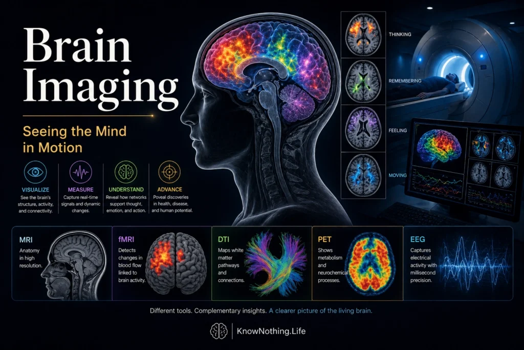
Structural imaging focuses on the physical anatomy of the brain, providing detailed information about its size, shape, and organization. MRI is the most widely used structural imaging technique, offering high-resolution images that can reveal subtle differences in brain tissue. It is particularly useful for identifying abnormalities such as tumors, lesions, and atrophy associated with neurodegenerative diseases.
Diffusion tensor imaging (DTI), a specialized form of MRI, allows researchers to map the brain’s white matter pathways by tracking the movement of water molecules along axons. This technique provides insights into connectivity, revealing how different regions of the brain are linked. Understanding these connections is crucial for studying conditions that involve disrupted communication between regions, such as traumatic brain injury or schizophrenia.
Structural imaging has also contributed to the study of normal variation in brain anatomy. Differences in cortical thickness, volume, and connectivity have been associated with cognitive abilities, personality traits, and developmental stages. However, interpreting these differences requires caution, as correlation does not imply causation, and individual variability is substantial.
Functional Imaging Techniques
Functional imaging techniques provide insight into how the brain operates during specific tasks or states. Functional MRI (fMRI) is one of the most widely used methods, measuring changes in blood oxygenation that occur when neural activity increases. This “blood-oxygen-level-dependent” (BOLD) signal allows researchers to identify regions that are active during particular cognitive processes, such as memory, attention, or language.
Positron emission tomography (PET) offers another approach, using radioactive tracers to measure metabolic activity or the distribution of specific neurotransmitters. PET scans have been particularly valuable in studying disorders such as Alzheimer’s disease, where changes in metabolism can be detected before structural damage becomes apparent.
Electroencephalography (EEG) and magnetoencephalography (MEG) provide complementary information by measuring electrical and magnetic activity in the brain. These techniques offer high temporal resolution, capturing rapid changes in neural activity, though with less spatial precision than fMRI. Together, these methods provide a more complete picture of brain function, combining information about where and when activity occurs.
Brain Imaging and Cognitive Neuroscience
Brain imaging has played a central role in the development of cognitive neuroscience, a field that seeks to understand the neural basis of mental processes. By linking patterns of brain activity to specific tasks, researchers can identify the networks involved in functions such as perception, memory, and decision-making.
For example, studies using fMRI have shown that memory involves interactions between the hippocampus and other cortical regions, while attention engages networks in the frontal and parietal lobes. These findings support the idea that cognitive functions are distributed across networks rather than localized in single regions. As Antonio Damasio argued in Descartes’ Error, cognition and emotion are deeply intertwined, a conclusion supported by imaging studies that reveal overlapping neural circuits.
Brain imaging has also contributed to the study of consciousness, providing insights into the neural correlates of awareness. While the nature of consciousness remains a subject of debate, imaging studies have identified patterns of activity associated with different states, such as wakefulness, sleep, and anesthesia. These findings offer a window into one of the most fundamental aspects of human experience.
Clinical Applications
In clinical settings, brain imaging is an essential tool for diagnosis and treatment planning. Structural imaging can identify physical abnormalities, while functional imaging can reveal patterns of activity associated with specific disorders. For example, reduced activity in certain brain regions has been observed in depression, while altered connectivity patterns are associated with schizophrenia.
Brain imaging is also used to monitor the progression of neurological diseases and evaluate the effectiveness of treatments. In conditions such as Alzheimer’s disease, imaging can detect early changes in brain structure and function, allowing for earlier intervention. Similarly, imaging can guide surgical procedures, helping clinicians avoid critical areas and improve outcomes.
Neurologist Oliver Sacks emphasized the importance of combining imaging with clinical observation, noting that “the neurological patient is not just a collection of deficits but a person with a story.” Imaging provides valuable data, but it must be interpreted within the context of the individual’s experience and history.
Limitations and Ethical Considerations
Despite its power, brain imaging has limitations. Functional imaging methods such as fMRI provide indirect measures of neural activity, relying on changes in blood flow rather than direct observation of neurons. This can lead to challenges in interpretation, particularly when trying to infer complex mental states from patterns of activity.
There is also a risk of overinterpretation, sometimes referred to as “neurorealism,” where brain images are seen as definitive evidence of psychological phenomena. While imaging can reveal correlations between brain activity and behavior, it does not necessarily explain causation. As a result, findings must be interpreted carefully, with an understanding of their limitations.
Ethical issues also arise in the use of brain imaging. Questions about privacy, consent, and the potential misuse of data are increasingly important as imaging technologies become more advanced. For example, the possibility of using brain imaging to infer thoughts or intentions raises concerns about autonomy and confidentiality.
Conclusion
Brain imaging has transformed the study of the mind, providing unprecedented access to the structure and function of the living brain. By revealing the neural basis of cognition, emotion, and behavior, it has deepened our understanding of human experience and advanced both research and clinical practice.
At the same time, brain imaging is not a complete solution to the mysteries of the mind. It offers powerful tools, but these must be integrated with other approaches to fully understand the complexity of human behavior. As the field continues to evolve, brain imaging will remain a central component of neuroscience, illuminating the intricate processes that underlie thought, feeling, and action.