
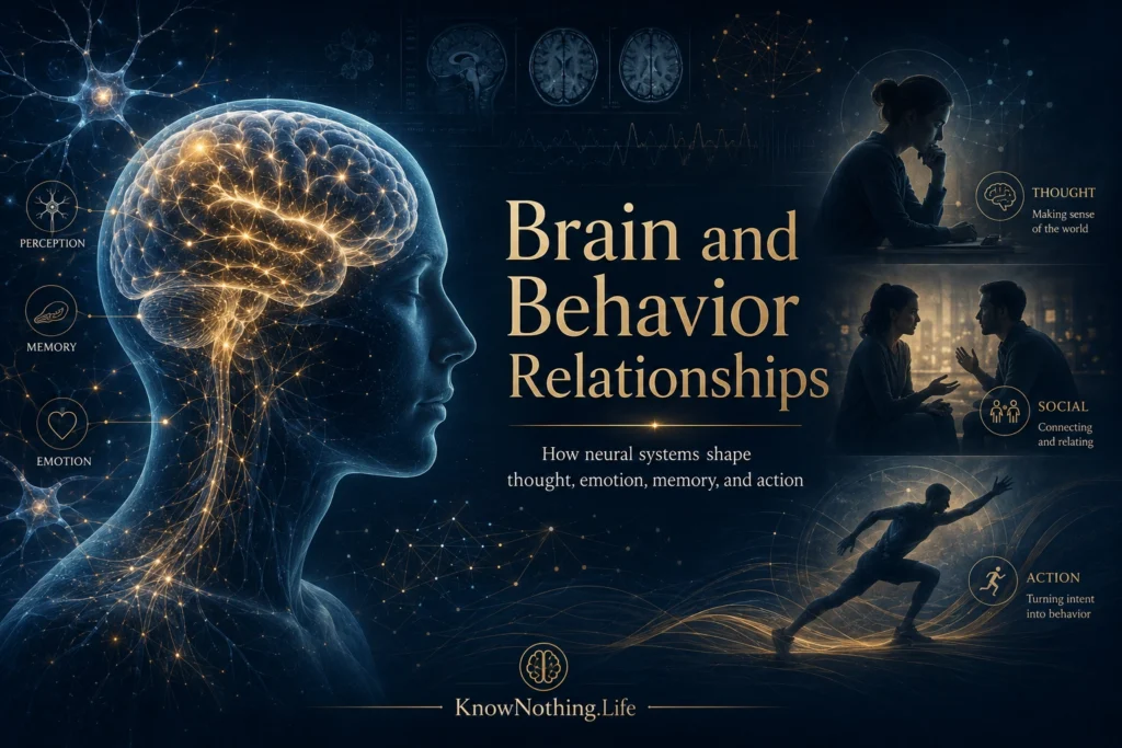
Brain and behavior relationships describe how activity within the nervous system contributes to perception, thought, emotion, memory, motivation, movement, and social conduct. Researchers ask how injury changes behavior, how neural activity accompanies mental processes, and how experience reorganizes the brain. The goal is not merely to assign each behavior to a location, but to explain how biological systems make meaningful action possible.
A complete explanation must operate at several levels. Fear, for example, involves sensory detection, learned associations, bodily arousal, attention, memory, and decisions about whether to approach or avoid a situation. These components depend on interactions among the amygdala, hippocampus, hypothalamus, brainstem, prefrontal cortex, and the rest of the body. Brain–behavior relationships are therefore both localized and distributed: particular structures make specialized contributions, while complex behavior emerges from coordinated networks.
Localization, Brain Injury, and Neural Networks
Neurological injuries provided some of the earliest evidence that different brain regions make different contributions to behavior. The case of railway worker Phineas Gage became famous because a severe frontal injury was followed by reported changes in judgment, emotional regulation, and social conduct. In a 1994 Science study, Hanna Damasio and colleagues reconstructed the likely path of the tamping iron through Gage’s skull and concluded that the damage probably involved portions of both prefrontal cortices associated with decision-making and emotion.
Cases such as Gage encouraged the localization of function, but they also revealed its limits. Lesions disrupt connections as well as local tissue, and the same injury can affect several psychological processes. Marcel Mesulam’s influential 1990 paper, “Large-Scale Neurocognitive Networks and Distributed Processing for Attention, Language, and Memory,” argued that complex behavior is supported by interconnected systems rather than single anatomical centers. His model helped establish the modern view that functions such as attention, memory, and language depend on specialized nodes working within distributed networks.
Hemispheric Specialization and Cooperation
Research involving people whose corpus callosum had been surgically divided offered dramatic evidence of hemispheric specialization. In 1962, Michael Gazzaniga, Joseph Bogen, and Roger Sperry reported that information presented to one visual field could sometimes guide behavior without being verbally identified. Because visual information reaches opposite hemispheres, these findings showed that perception, language, and action could be partly separated when communication between the hemispheres was interrupted.
The results are often distorted into the claim that individuals are either “left-brained” or “right-brained.” Ordinary behavior depends on cooperation between hemispheres. Language may be strongly left-lateralized in many people, while aspects of spatial attention and emotional communication involve greater right-hemisphere participation. Reading, planning, and social understanding nevertheless require bilateral networks. Specialization increases efficiency, but integration produces coherent experience.
Memory Systems and the Hippocampus
One of the clearest demonstrations of a brain–behavior relationship came from William Scoville and Brenda Milner’s 1957 report on patients who had undergone bilateral medial temporal lobe surgery. Their most extensively studied patient, later publicly identified as Henry Molaison, developed profound difficulty forming new long-term declarative memories while retaining short-term information and some forms of skill learning. The findings showed that memory is not a single ability and that medial temporal structures, including the hippocampus, are especially important for creating new memories of facts and events.
Molaison improved on certain motor tasks even though he did not consciously remember practicing them, helping distinguish procedural learning from declarative memory. Later research showed that memory depends on interactions among the hippocampus, cortex, amygdala, basal ganglia, and cerebellum. Different systems contribute to autobiographical memory, emotional learning, habits, working memory, navigation, and motor skills.
Perception, Action, and the Construction of Experience
Vision appears effortless, yet it depends on multiple transformations across the retina, thalamus, visual cortex, and association areas. David Hubel and Torsten Wiesel’s classic recordings from the cat visual cortex showed that individual neurons respond selectively to features such as line orientation and position. Their 1959 study helped establish that perception is actively constructed through organized neural processing rather than delivered to consciousness as an unchanged picture of the outside world.
The same principle applies to touch, hearing, and movement. Sensory systems represent the body and environment, while motor systems select goals, coordinate muscles, compare feedback with predictions, and correct errors. Michael Merzenich and colleagues found that the somatosensory cortical map reorganized after digit amputation in adult monkeys, demonstrating that mature sensory cortex retains substantial plasticity. Such reorganization supports adaptation, although it may also contribute to phantom sensations and chronic pain.
Emotion, Reward, and Decision-Making
Emotion is sometimes treated as the opposite of reason, but behavioral neuroscience shows that the two are deeply connected. Joseph LeDoux and his colleagues traced pathways through which auditory information reaches the amygdala during fear conditioning. Their research demonstrated how a previously neutral cue can acquire the ability to trigger defensive responses after being paired with danger. The amygdala does not contain fear as a single experience; it participates in networks linking perception with arousal, attention, memory, and action.
Research on reward and decision-making produced a similar lesson. James Olds and Peter Milner reported in 1954 that rats repeatedly pressed a lever to receive electrical stimulation in certain brain regions, showing that neural activity could serve as a powerful reinforcer. Four decades later, Antoine Bechara and colleagues found that patients with prefrontal damage repeatedly favored immediate gains despite larger future losses on a gambling task. Together, these studies showed that motivation and choice depend on systems that integrate reward, emotion, learning, and anticipated consequences.
Plasticity and the Influence of Experience
The brain changes in response to experience throughout life. Learning can alter synaptic strength, patterns of connectivity, gene expression, and the organization of functional networks. Plasticity allows people to acquire language, develop skills, adapt to new environments, and recover some abilities after injury. It also means that repeated stress, avoidance, substance use, or compulsive behavior can strengthen patterns that become increasingly difficult to change.
Plasticity is powerful but not unlimited. Developmental timing, genetics, age, injury severity, sleep, and social support influence what change is possible. The nervous system must balance adaptability with stability; a brain that reorganized completely after every event could not preserve reliable memories or skills. Brain–behavior relationships are dynamic but constrained by biology and experience.
How Researchers Establish Cause and Effect
Scientists investigate brain–behavior relationships through lesion analysis, electrophysiology, neuroimaging, brain stimulation, pharmacology, genetics, and carefully designed behavioral tasks. Functional magnetic resonance imaging can identify changes in blood oxygenation associated with neural activity, but it measures a vascular response and is usually correlational. A brightly colored region on a scan does not prove that the region uniquely creates the behavior under investigation.
Causal methods offer stronger tests. In 2005, Edward Boyden and colleagues demonstrated millisecond-scale optical control of genetically targeted neurons, helping establish optogenetics as a method for activating selected cell populations and observing resulting behavior in laboratory animals. Even this technique requires caution because artificial stimulation may not reproduce natural activity, and animal models cannot capture every dimension of human experience. Strong conclusions usually come from several methods producing compatible evidence.
Clinical Meaning and the Whole Person
Knowledge of brain–behavior relationships guides the understanding and treatment of stroke, traumatic brain injury, epilepsy, Parkinson’s disease, dementia, addiction, depression, and anxiety. Neuropsychological testing reveals patterns of preserved and impaired ability, while rehabilitation uses remaining capacities and plasticity to restore function or develop compensatory strategies.
The brain is necessary for behavior, but behavior cannot be understood from the brain alone. Hormones, immune activity, bodily feedback, relationships, culture, language, and physical surroundings all influence neural function. Brain and behavior form a reciprocal system: neural activity produces action, action changes the environment, and environmental consequences reshape future neural activity. The strongest explanations therefore connect cells and circuits with personal history, social context, and the purposes that behavior serves.


