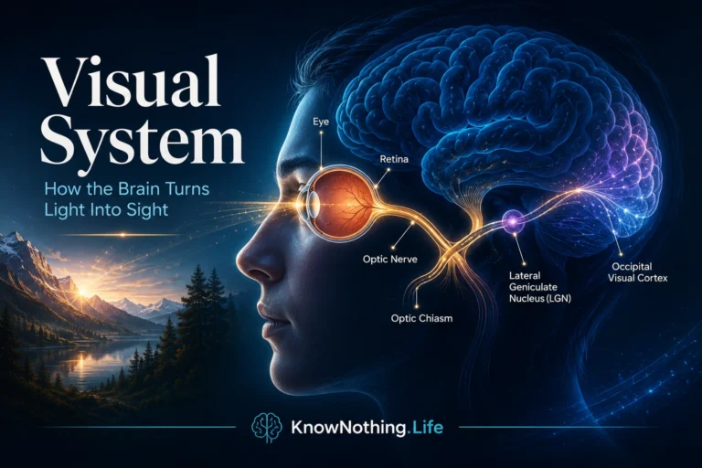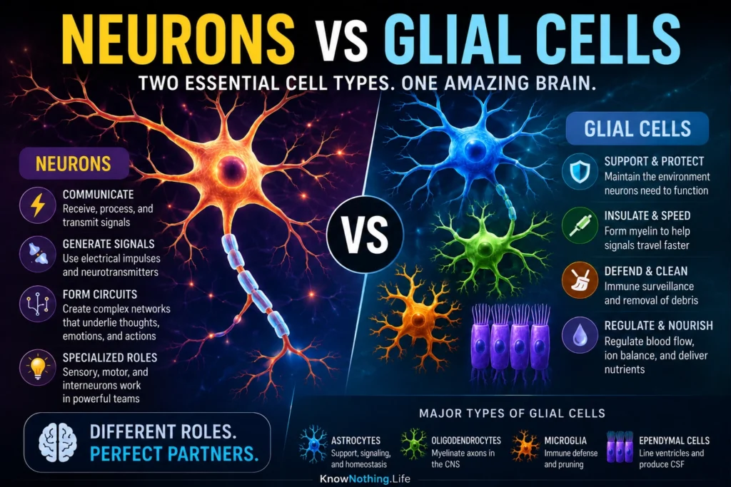
Neurons and glial cells are the two major cellular families that make up the nervous system. Neurons are the specialized signaling cells responsible for receiving, processing, and transmitting information through electrical and chemical activity. A typical neuron has a soma, or cell body, containing the nucleus, along with dendrites that receive input, an axon that sends signals, and synapses that communicate with other cells. This structure allows neurons to support sensation, movement, perception, memory, emotion, reflexes, and thought. NCBI’s StatPearls describes neurons as cells with a soma containing the nucleus, an axon, and a dendritic tree, with synapses forming points of communication between connected neurons.
Glial cells, often called neuroglia, are non-neuronal cells that support, regulate, protect, and help organize nervous-system function. The main glial cells in the central nervous system include astrocytes, oligodendrocytes, microglia, and ependymal cells, while Schwann cells and satellite cells serve major roles in the peripheral nervous system. Older textbooks sometimes described glia as “glue,” but that image is outdated. Glial cells do not simply hold neurons in place. They regulate the chemical environment, form myelin, support synapses, participate in immune defense, guide development, and help maintain the conditions that allow neurons to signal properly. NCBI’s Neuroscience text identifies astrocytes, oligodendrocytes, and microglial cells as major glial types in the mature central nervous system and describes astrocytes as helping maintain an appropriate chemical environment for neuronal signaling.
The Core Difference: Signaling vs Support and Regulation
The simplest comparison is that neurons are the main rapid signaling cells, while glial cells are the main support and regulatory cells. Neurons generate action potentials, release neurotransmitters, form synaptic circuits, and carry information across long distances. Sensory neurons carry information from the body toward the brain and spinal cord. Motor neurons carry commands from the central nervous system toward muscles. Interneurons connect neurons within local circuits and are especially abundant in the brain and spinal cord. Their basic design is built around communication: dendrites receive, the soma integrates, the axon transmits, and synapses influence the next cell.
Glial cells usually do not transmit information in the same fast action-potential-based way neurons do, but they are deeply involved in shaping how neuronal communication works. Astrocytes help regulate ions, neurotransmitters, metabolism, synapse formation, and the blood-brain barrier. Oligodendrocytes and Schwann cells form myelin, allowing axons to conduct signals faster and more efficiently. Microglia monitor the nervous system, respond to injury, and help remodel circuits. Ependymal cells line the ventricles and contribute to cerebrospinal fluid function. In other words, neurons may carry the main messages, but glia help determine whether those messages are possible, efficient, protected, and properly regulated.
Neurons: The Nervous System’s Communication Units
Neurons are built for excitability. They maintain ion gradients across their membranes, allowing them to respond to incoming signals and generate action potentials when threshold is reached. These electrical impulses travel down the axon and often trigger neurotransmitter release at synapses. A neuron can receive thousands of inputs and produce output that influences many other cells. This makes neurons the core communication units of the nervous system. NCBI’s Developmental Biology text summarizes neurons as cells that transmit electrical impulses from one region of the body to another, typically receiving signals through dendrites and focusing them toward the axon.
The importance of neurons was clarified through the neuron doctrine, the idea that the nervous system is made of individual cellular units rather than one continuous web. Santiago Ramón y Cajal and Camillo Golgi shared the 1906 Nobel Prize for their work on the structure of the nervous system, even though they disagreed about how nerve cells were organized. A historical review notes that Cajal and Golgi held opposing views, with the neuron doctrine emphasizing the structural, functional, and developmental individuality of nerve cells. This doctrine became one of the foundations of modern neuroscience because it made the neuron the basic unit for studying nervous-system function.
Glial Cells: The Nervous System’s Hidden Infrastructure
Glial cells are essential because neurons cannot function alone. A neuron needs energy, chemical stability, insulation, immune protection, waste clearance, synaptic regulation, and developmental support. Astrocytes are among the most important examples. They help regulate extracellular potassium, clear excess neurotransmitters, support metabolism, promote synapse formation, and contribute to blood-brain barrier function. StatPearls describes astrocytes as performing metabolic, structural, homeostatic, and neuroprotective tasks, including clearing excess neurotransmitters, stabilizing the blood-brain barrier, and promoting synapse formation.
Oligodendrocytes and Schwann cells show another major glial role: myelination. Oligodendrocytes form myelin in the central nervous system, while Schwann cells form myelin in the peripheral nervous system. Myelin wraps around axons and allows electrical signals to travel more rapidly through saltatory conduction. Research reviews emphasize that oligodendrocytes do more than wrap axons; they also support long-term axonal integrity and metabolic function. This means glia are not just passive insulation. They help keep neural pathways healthy and functional over time.
Astrocytes, Synapses, and the Tripartite Synapse
Astrocytes have changed how scientists think about synapses. The traditional model of a synapse includes a presynaptic neuron, a synaptic cleft, and a postsynaptic neuron. The tripartite synapse concept adds astrocytes as active participants in synaptic function. Astrocytes can surround synapses, detect neurotransmitter release, regulate extracellular chemistry, influence receptor activity, and help shape synaptic strength. A review on astrocytes and synaptic activity describes the tripartite synapse as a concept in which astrocytes are active elements of synaptic physiology.
This does not mean astrocytes replace neurons as the main signaling cells. Rather, they regulate the environment in which neuronal signaling happens. Astrocytes can influence whether neurotransmitters remain in the synaptic space, how strongly synapses respond, and how plastic synaptic connections become. Research on astrocytes and synaptic plasticity notes that astrocytes regulate synaptic transmission and participate in long-term potentiation and functional synaptic change. This makes the old neuron-only picture incomplete. Learning, memory, and circuit function depend not only on neurons firing, but also on glial cells helping regulate the synaptic landscape.
Microglia, Immunity, and Synaptic Pruning
Microglia are often described as the immune cells of the central nervous system. They monitor the brain and spinal cord, respond to injury or disease, remove debris, and participate in inflammation. But microglia are not only emergency responders. They also help shape neural circuits during development and plasticity. One of their most important roles is synaptic pruning, the process by which unnecessary or weak synapses are removed so circuits become more efficient and refined.
Research on complement proteins and microglia has shown that immune-related mechanisms help tag and eliminate synapses during development. A review on complement and synaptic pruning states that complement proteins are localized to developing central-nervous-system synapses during periods of active synapse elimination and are required for normal brain wiring. Later reviews describe microglial synaptic pruning as a regulated process that begins in development and can also occur in other contexts. This means glial cells help sculpt the nervous system, not merely protect it after damage.
Cell Numbers and the Old Glia Myth
For a long time, people often claimed that glial cells outnumber neurons by ten to one. Modern cell-counting research has challenged that simplified statement. Azevedo and colleagues used the isotropic fractionator method and estimated that the adult male human brain contains about 86.1 billion neurons and about 84.6 billion non-neuronal cells. A review by von Bartheld and colleagues explains that the older belief in a 10:1 glia-to-neuron ratio has been challenged by newer methods, which suggest a ratio much closer to 1:1 in the human brain as a whole.
This correction matters because it prevents two opposite mistakes. The first mistake is treating neurons as the only important brain cells. The second is treating glia as mysterious background cells whose numbers alone explain brain power. The better view is relational. Neurons and glial cells exist in roughly comparable numbers in the human brain overall, but their ratios vary by region, species, development, and cell type. What matters most is not only how many cells exist, but how they interact. Nervous-system function emerges from neuron-glia partnerships.
Neurons and Glia in Disease
Many neurological and psychiatric conditions involve both neurons and glial cells. Neuronal dysfunction is central to epilepsy, neurodegenerative disease, stroke, traumatic brain injury, movement disorders, and many sensory or motor deficits. But glial dysfunction can be just as important. Multiple sclerosis involves damage to myelin in the central nervous system, making oligodendrocytes and myelin central to the disease process. Astrocyte dysfunction can affect blood-brain barrier regulation, neurotransmitter clearance, inflammation, and scar formation after injury. Microglia can help protect the brain, but excessive or misdirected inflammatory activity may contribute to disease processes.
This is why modern neuroscience increasingly studies neuron-glia networks rather than neurons alone. A disorder may begin with abnormal neuronal firing, altered glial support, immune activation, myelin damage, metabolic stress, or synaptic dysfunction. Once one part of the system is disturbed, the others may change as well. The brain is not a collection of isolated cell types. It is a living tissue ecosystem in which signaling, support, immunity, metabolism, and plasticity are tightly connected.
Why Neurons vs Glial Cells Matters
The comparison between neurons and glial cells matters because it clarifies how the nervous system works. Neurons are the main rapid communication cells. They fire action potentials, release neurotransmitters, and form circuits that support sensation, movement, memory, and thought. Glial cells are the essential regulators and support systems. They maintain the chemical environment, form myelin, guide development, influence synapses, protect against injury, regulate inflammation, and help keep neural circuits functional.
The deeper lesson is that the brain is not only a network of firing neurons. It is a cellular community. Neurons provide the electrical and chemical signals most directly associated with information processing, but glial cells make those signals reliable, efficient, protected, and adaptable. To understand neurons vs glial cells is to understand that brain function depends on both message and maintenance, signal and support, communication and regulation. The mind emerges not from neurons alone, but from the coordinated life of many kinds of brain cells working together.