
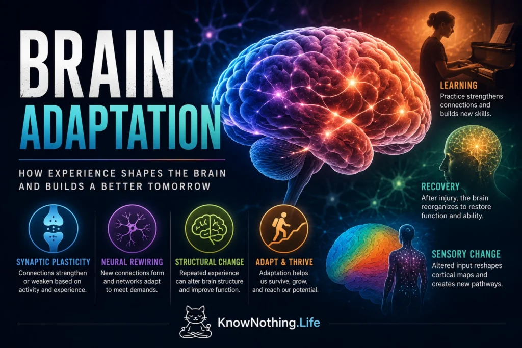
Brain adaptation is the nervous system’s ability to alter its activity, organization, and physical structure in response to experience, practice, injury, and changing environmental demands. The term includes several processes commonly grouped under neuroplasticity. Synapses can become stronger or weaker, neurons can change their excitability, dendritic spines can be remodeled, and cortical maps can shift according to patterns of use. Adaptation occurs most rapidly during development, but it does not end in adulthood. Research on sensory maps, motor learning, and structural brain change shows that the mature nervous system retains a constrained but meaningful ability to revise how information is represented and how behavior is produced.
This flexibility allows the brain to learn skills, improve perception, compensate for altered sensory input, and recover some functions after damage. Yet adaptation is not automatically beneficial. The same capacity that supports expertise and rehabilitation can also contribute to chronic pain, phantom sensations, or inefficient compensatory patterns. Brain adaptation is therefore better understood as a biological response to repeated demands than as an automatic process of improvement. Neural circuits become increasingly suited to the activity, attention, and sensory information they repeatedly encounter, whether or not the resulting pattern serves a person’s long-term interests.
Synaptic Change and Homeostatic Balance
At the smallest functional scale, adaptation begins with changes at synapses, the junctions through which neurons influence one another. Repeated patterns of activity can strengthen transmission through long-term potentiation or weaken it through long-term depression. These changes involve neurotransmitter receptors, calcium-dependent signaling, protein modification, gene expression, and sometimes the growth or elimination of dendritic spines. Synaptic plasticity gives experience a way to alter how strongly one neuron affects another, allowing coordinated signals to gain influence while poorly timed or less useful inputs are reduced. Changes occurring across large numbers of synapses can eventually modify the behavior of entire networks.
Adaptation also requires stability. If every active connection strengthened without limit, neural networks could become saturated or excessively excitable. Gina Turrigiano and colleagues demonstrated a compensatory process called synaptic scaling in their 1998 study Activity-Dependent Scaling of Quantal Amplitude in Neocortical Neurons. When cultured cortical neurons experienced prolonged reductions in activity, their excitatory synapses strengthened broadly. Increased network activity produced scaling in the opposite direction. Unlike input-specific learning mechanisms, synaptic scaling adjusts many connections while preserving their relative differences, helping neurons remain responsive without losing previously encoded information. Brain adaptation therefore depends on a continuing balance between plastic change and homeostatic regulation.
Experience Reshapes Cortical Maps
One of the clearest demonstrations of adult brain adaptation came from experiments showing that cortical maps are not permanently fixed. Michael Merzenich and colleagues examined the somatosensory cortex of adult monkeys after restricted loss of sensory input. In their 1983 study Topographic Reorganization of Somatosensory Cortical Areas 3b and 1 in Adult Monkeys Following Restricted Deafferentation, cortical regions that had previously represented the affected skin gradually became responsive to neighboring body surfaces. The result challenged the idea that mature sensory maps were rigidly assigned. It showed that surviving inputs can compete for cortical territory when the pattern of information arriving from the body changes.
Training can produce similar reorganization without injury. William Jenkins and colleagues trained adult owl monkeys on demanding tactile tasks and found that representations of the practiced skin surfaces expanded in the primary somatosensory cortex. Randy Nudo and colleagues later trained squirrel monkeys on skilled hand movements. Movement representations used during successful training grew within the primary motor cortex, while less-used representations contracted. The changes were progressive and related to skill acquisition rather than movement repetition alone. The brain adapted because the animals had to use sensory feedback and precise movements to solve a behavioral problem, demonstrating that the quality and relevance of practice influence cortical reorganization.
Structural Changes in the Human Brain
Human imaging studies indicate that learning can be accompanied by measurable structural change. In 2004, Bogdan Draganski and colleagues studied adults learning to juggle and reported temporary increases in gray matter in regions associated with processing visual motion. Participants were scanned before learning, after acquiring the skill, and again after a period without practice. Some of the detected changes diminished when training stopped. Magnetic resonance imaging could not reveal the exact cellular process responsible, so the results did not prove that new neurons had appeared. They nevertheless demonstrated that acquiring a novel skill can produce measurable alterations in adult brain anatomy over a period of months.
Eleanor Maguire and colleagues found another striking association in licensed London taxi drivers, who were required to acquire extensive knowledge of the city’s streets and landmarks. Their 2000 study reported greater relative volume in the posterior hippocampus of taxi drivers than in control participants, while a more anterior hippocampal region was larger in controls. A later comparison with London bus drivers helped separate navigation demands from driving experience and occupational stress. Taxi drivers again showed greater gray matter volume in mid-posterior hippocampal regions. These findings suggest that prolonged demands on spatial memory are associated with regionally specific structural adaptation, although observational studies cannot prove that experience caused every anatomical difference.
Adaptation to Sensory Loss
When ordinary sensory input is unavailable, cortical regions may become responsive to information from other senses. Norihiro Sadato and colleagues used functional brain imaging to study Braille reading in people who had been blind from an early age. Tactile discrimination and Braille tasks activated primary and secondary visual cortical regions, whereas the same regions did not respond in the same way among sighted controls. The visual cortex was therefore participating in the processing of touch rather than remaining inactive simply because visual information was absent.
This cross-modal organization shows that the function of a cortical region is shaped not only by its anatomical location but also by the information available to it and the tasks a person repeatedly performs. The occipital cortex remains connected to wider brain networks capable of carrying attention, language, memory, and spatial information. When visual input is absent, some of these connections may become functionally more influential. Such adaptation does not mean that every region can perform any possible task. Existing anatomy still creates limits, but experience can alter how available networks are recruited and coordinated.
Brain Adaptation After Injury
Adaptation is central to rehabilitation following neurological injury. Nudo and colleagues examined monkeys that had experienced small injuries to the part of the motor cortex controlling the hand. Animals receiving focused rehabilitative training recovered skilled movement more successfully, and surviving cortical tissue near the damaged region developed altered movement representations. Without comparable training, representation of the affected hand could shrink further. The findings suggested that rehabilitation does more than strengthen muscles or teach conscious strategies. Repeated, meaningful practice can influence how surviving brain tissue reorganizes after injury.
Human studies of constraint-induced movement therapy have reported related patterns after stroke. This approach limits reliance on the less-affected arm while requiring intensive practice with the affected limb. Studies by Edward Taub, Joachim Liepert, and other researchers found improved motor performance together with changes in the cortical representation of the recovering limb. Recovery does not usually mean that the brain returns perfectly to its earlier condition. Surviving networks may become stronger, previously weak pathways may be recruited, and behavior may be reorganized around remaining capacity. The type, timing, repetition, and difficulty of rehabilitation help determine which compensatory patterns become established.
The Limits and Costs of Plasticity
The brain’s capacity to adapt is substantial but constrained. Plasticity depends on developmental stage, existing connections, the location and severity of injury, and the patterns of activity available to guide change. Adult cortical representations can reorganize, but the process does not make the brain infinitely rewritable. Some lost functions may be only partly recovered because the original cells, pathways, or developmental conditions cannot be recreated. Increased activation on a brain scan is also not automatically evidence of improvement. It may indicate efficient specialization, greater effort, compensation by another region, or an unstable phase of recovery.
Adaptation can also become maladaptive. Herta Flor and colleagues found a close relationship between somatosensory cortical reorganization and phantom-limb pain after amputation. Later research connected altered cortical representation with other persistent pain conditions. These findings suggest that repeated pain-related signaling can become embedded in the organization of sensory networks, allowing discomfort to continue even when ordinary tissue healing cannot fully explain it. Plasticity’s fundamental rule is therefore neutral: the brain changes according to repeated activity and behavioral importance, not according to an independent definition of health.
Why Brain Adaptation Matters
Brain adaptation explains how the nervous system can remain stable enough to preserve knowledge and identity while changing enough to learn. It operates across several levels. Receptors move into or out of synapses, gene expression changes cellular behavior, dendritic spines appear or disappear, cortical representations compete for territory, and distributed networks develop new ways of coordinating perception and action. No single mechanism accounts for every form of adaptation. What unites these processes is their ability to convert repeated experience into a lasting alteration in future neural processing.
The evidence replaces two misleading extremes. The adult brain is neither permanently fixed nor endlessly malleable. It is a living system whose capacity for change is powerful, selective, and dependent on conditions. Practice can reshape sensory and motor representations, sustained cognitive demands can be associated with structural differences, altered input can recruit new cortical resources, and rehabilitation can guide surviving circuits after injury. At the same time, plasticity has limits and can preserve harmful patterns as readily as useful ones. Understanding brain adaptation therefore means recognizing both possibility and constraint: experience changes the brain, but the direction and durability of that change depend greatly on how experience is structured.


