
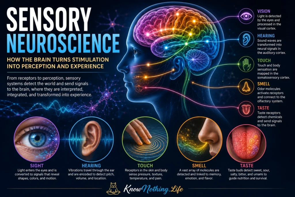
Sensory neuroscience is the study of how nervous systems detect events within the body and surrounding environment, convert them into neural signals, and organize those signals into perception and behavior. It encompasses the familiar senses of vision, hearing, smell, taste, and touch, but it also includes balance, temperature, pain, itch, proprioception, and internal sensations arising from organs. These systems allow the brain to estimate what is happening, where it is occurring, how important it may be, and what response is appropriate.
Sensation is not a direct recording of physical reality. Sensory receptors respond selectively to particular forms of energy or chemical stimulation, and neural circuits preserve some features while transforming or discarding others. The result is an efficient but incomplete representation shaped by receptor sensitivity, neural organization, attention, previous experience, and behavioral needs. Sensory neuroscience therefore investigates both the biological machinery that receives information and the computations through which the brain turns that information into a meaningful world.
Sensory Transduction and Neural Coding
Every sensory system begins with transduction: the conversion of physical or chemical energy into electrical activity. Photoreceptors respond to light, cochlear hair cells respond to mechanical vibration, olfactory receptors bind airborne molecules, taste receptors detect dissolved compounds, and somatosensory receptors respond to pressure, temperature, tissue damage, or body movement. These receptors alter ion flow across their membranes, producing electrical signals that can influence the firing of sensory neurons. Transduction allows fundamentally different stimuli to enter the nervous system through a shared language of changing membrane potentials and action potentials.
The brain must then determine what a pattern of neural activity represents. Information may be encoded through which neurons respond, how rapidly they fire, when they fire relative to other cells, and how activity is distributed across a population. Sensory neurons usually possess receptive fields, meaning that each responds most strongly to stimulation within a particular region or range. The receptive-field concept was developed through early studies of touch and vision and later became central to nearly every sensory field. It provided a way to connect measurable neural responses with specific features of an organism’s environment.
Touch, Proprioception, and the Somatosensory System
The somatosensory system includes several related but distinct capacities. Mechanoreceptors in the skin respond to indentation, vibration, stretching, and movement across the body surface. Thermoreceptors signal warming and cooling, while nociceptors respond to potentially damaging mechanical, chemical, or thermal events. Proprioceptors in muscles, tendons, and joints provide information about limb position and movement. Together, these systems allow people to recognize an object by touch, maintain posture, coordinate movement, detect injury, and know where their bodies are without continuously looking at them.
Mechanical sensation depends partly on PIEZO2, an ion channel that opens in response to physical force. Mouse studies identified PIEZO2 as a major transducer for touch and a principal channel for proprioception, while research involving people with inactivating PIEZO2 variants demonstrated selective impairments in discriminative touch and awareness of body position. Somatosensory information also retains an organized representation in the cortex. Vernon Mountcastle’s recordings from somatosensory cortex helped establish that vertically arranged neurons could share receptive-field and modality properties, influencing later ideas about functional cortical organization.
Vision and the Organization of Visual Information
Vision begins when light reaches photoreceptors in the retina, but substantial processing occurs before information leaves the eye. Retinal circuits compare signals across space, emphasize contrast, and separate aspects of visual input into parallel pathways. Retinal ganglion cells transmit the resulting activity through the optic nerve to several brain structures, including the lateral geniculate nucleus of the thalamus and the superior colliculus. Signals reaching primary visual cortex remain spatially organized, creating a retinotopic map in which neighboring cortical locations generally represent neighboring portions of the visual field.
David Hubel and Torsten Wiesel transformed visual neuroscience by recording from individual neurons in the visual cortex. In their 1962 study of the cat visual system, they showed that many cortical neurons respond selectively to edges or bars presented at particular orientations and positions. They distinguished simple and complex receptive fields and described an organized cortical architecture involving orientation and ocular preference. Later work in monkeys found comparable response properties, helping establish the idea that visual perception emerges through successive neural transformations rather than from one internal picture reproduced inside the brain.
Hearing and the Neural Analysis of Sound
Hearing begins when sound waves vibrate the eardrum and middle-ear bones, transferring mechanical energy into the fluid-filled cochlea. Movement along the cochlear partition bends sensory hair cells, causing them to alter neurotransmitter release onto auditory nerve fibers. Different locations along the cochlea respond most strongly to different sound frequencies, producing a tonotopic arrangement. This frequency-based organization is preserved through much of the ascending auditory system, although later neural processing also extracts timing, intensity, spatial location, pitch, speech patterns, and the acoustic features of voices or environmental sounds.
Evidence for tonotopic organization in human auditory cortex emerged from neuromagnetic recordings by Gian Luca Romani, Samuel Williamson, and Lloyd Kaufman. Their 1982 study found that the estimated cortical source of responses shifted systematically with sound frequency. Subsequent fMRI research has mapped multiple frequency gradients across the auditory cortex, while also showing that physical frequency and perceived pitch do not always share identical representations. Hearing therefore depends on both orderly sensory maps and more complex transformations that allow the brain to recognize meaningful sound patterns.
Smell, Taste, and Chemical Sensing
Olfaction begins when odor molecules interact with receptors on sensory neurons in the nasal epithelium. Linda Buck and Richard Axel’s 1991 paper, “A Novel Multigene Family May Encode Odorant Receptors,” identified a large family of genes that appeared to encode odorant receptors. Their discovery provided a molecular explanation for how a limited number of receptor types could distinguish an enormous range of odors. Individual odorants can activate combinations of receptors, allowing smells to be represented through distributed patterns rather than requiring one receptor for every possible substance.
Taste uses several receptor mechanisms to identify chemicals that may signal nutrients, minerals, toxins, or acidity. T1R receptor combinations contribute to sweet and umami detection, whereas the T2R family is strongly associated with bitter compounds. Experimental disruption of signaling molecules such as TRPM5 or PLCβ2 can eliminate normal responses to sweet, bitter, and umami stimuli in mice while leaving other taste categories less affected. Flavor nevertheless depends on more than taste receptors alone. Smell, texture, temperature, irritation, expectation, and visual information combine to create the experience associated with food and drink.
Multisensory Integration and Perceptual Unity
Sensory systems are often studied separately, but natural perception requires them to cooperate. Seeing a speaker’s face can change how speech sounds are interpreted, while a sudden sound can redirect visual attention toward its apparent location. Studies of neurons in the superior colliculus showed that visual, auditory, and somatosensory responses can converge within individual cells. Meredith and Stein found that multisensory enhancement depends on spatial and temporal relationships: stimuli that occur near one another in location and time are more likely to be combined than signals suggesting unrelated events.
Integration does not mean that the senses lose their specialized organization. Instead, sensory information is exchanged across cortical and subcortical networks while maintaining features needed for specific tasks. The brain must decide whether signals share a common cause, such as whether a voice belongs to a visible person or a vibration accompanies a touched object. These computations help produce perceptual unity, allowing several streams of neural activity to be experienced as one coherent event rather than as unrelated fragments.
Plasticity, Sensory Loss, and the Future of the Field
Sensory circuits remain capable of change throughout life. Developmental experience helps refine receptive fields and cortical maps, while training can improve discrimination of sounds, textures, movements, or visual features. Following sensory loss, regions usually associated with one modality may participate in processing information delivered through another. Studies of congenitally blind adults trained with visual-to-auditory sensory-substitution systems have found recruitment of occipital regions during tasks involving shapes, bodies, or written symbols conveyed through sound. These findings suggest that some cortical areas may be organized partly around computations rather than being permanently restricted to one sensory input.
Future sensory neuroscience will increasingly combine cellular recordings, brain imaging, genetics, computational modeling, virtual environments, and neural interfaces. These methods may improve cochlear and retinal prostheses, treatments for chronic pain, rehabilitation after sensory loss, and devices that translate information between modalities. Yet the field’s central challenge remains unchanged: explaining how electrochemical activity becomes the felt qualities of color, sound, pressure, pain, flavor, and bodily presence. Sensory neuroscience reveals that perception begins at specialized receptors, but it becomes meaningful only through the coordinated activity of the brain.


