
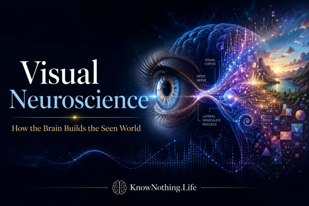
Visual neuroscience studies how the eyes and brain transform light into representations of objects, movement, depth, color, faces, places, and spatial relationships. Vision feels immediate, as though the world simply appears before consciousness, but the nervous system receives only changing patterns of light projected onto two retinal surfaces. From these signals, it must estimate what is present, distinguish objects from backgrounds, compensate for movement, identify relevant details, and guide behavior. The field therefore examines not only eyesight but also the neural computations through which coherent visual experience is constructed.
Visual processing is distributed across interconnected structures rather than confined to a single visual center. The retina begins analyzing information before signals leave the eye. Activity then travels through the optic nerve to targets including the lateral geniculate nucleus of the thalamus, the superior colliculus, and other subcortical structures. Information supporting detailed vision reaches primary visual cortex, or V1, before spreading through numerous extrastriate areas. Feedforward, lateral, and feedback connections continually reshape visual activity according to context, attention, expectation, and action.
The Retina as a Neural Processor
The retina is a layered part of the central nervous system containing photoreceptors, bipolar cells, horizontal cells, amacrine cells, and retinal ganglion cells. Rods are highly sensitive under dim illumination, while cones support daylight vision, color discrimination, and fine spatial detail. Photoreceptors do not send an untouched copy of the retinal image toward the brain. Local circuits compare signals across neighboring regions, adjust sensitivity to illumination, detect changes over time, and divide information into parallel channels. The axons of retinal ganglion cells form the optic nerve, making their patterns of activity the retina’s principal output.
Stephen Kuffler’s 1953 study, “Discharge Patterns and Functional Organization of Mammalian Retina,” helped establish the receptive-field concept. Kuffler showed that retinal ganglion cells respond according to the spatial arrangement of light within center and surrounding regions. Some cells are excited by illumination in the receptive-field center and inhibited by illumination in the surround, while others display the reverse organization. These center-surround relationships emphasize contrast and boundaries rather than absolute brightness. A nearly uniform surface may consequently generate less informative activity than an edge, shadow, or sudden change in illumination.
From the Thalamus to Primary Visual Cortex
Retinal signals partially cross at the optic chiasm, allowing information from the left visual field to be processed predominantly by the right cerebral hemisphere and information from the right field by the left. Many retinal axons terminate in the lateral geniculate nucleus, where signals from the two eyes and different retinal pathways remain organized. The thalamus is not merely a passive relay station. Its activity is influenced by cortical feedback, arousal, attention, and behavioral state, enabling the brain to regulate the flow of visual information before it reaches V1.
David Hubel and Torsten Wiesel’s recordings from the cat visual cortex produced one of neuroscience’s most influential accounts of cortical feature selectivity. Their 1962 study found neurons that responded strongly to bars or edges presented at particular orientations and positions. They distinguished simple and complex receptive fields, documented binocular interactions, and described an orderly cortical architecture. Later experiments demonstrated similar principles in primates. These findings showed that V1 does not function as an internal screen. It transforms retinal and thalamic input into representations of contours, orientation, spatial frequency, direction, and binocular disparity.
Visual Maps and the Perception of Depth
Primary visual cortex contains a retinotopic map in which neighboring points in the visual field are generally represented by neighboring cortical regions. The map is distorted rather than geometrically uniform because central vision receives disproportionately extensive cortical representation. This cortical magnification corresponds to the high spatial resolution of the fovea. Each hemisphere’s V1 mainly represents the opposite half of visual space, while the upper and lower visual fields occupy different regions around the calcarine sulcus. Experimental mapping has demonstrated this orderly representation across primate visual cortex.
The brain must also reconstruct depth from two-dimensional retinal images. Because the eyes view the world from slightly different positions, corresponding features fall at different retinal locations. Neurons sensitive to binocular disparity use these differences to support stereoscopic depth perception. Perspective, shading, relative size, texture, occlusion, and motion parallax provide additional information. No single cue offers a perfect measurement under every condition. Perceived depth emerges as the nervous system combines several signals according to their probable reliability.
Motion, Color, and Functional Specialization
Visual information leaving V1 reaches multiple extrastriate regions with different response preferences. Area MT, also called V5, contains many neurons selective for the direction and speed of movement. In 1988, William Newsome and Edward Paré found that lesions involving MT selectively impaired monkeys’ ability to discriminate motion direction within affected portions of the visual field. Human neuroimaging later identified a motion-responsive cortical region with comparable properties. These findings support an important role for MT in visual motion while also showing that motion perception depends on a broader network of cortical areas.
Color vision begins with comparisons among different cone classes, but stable color perception requires increasingly complex cortical computations. The wavelength reaching the eye changes dramatically with illumination, yet surfaces often retain approximately constant perceived colors. Semir Zeki’s work helped establish the principle of functional specialization, and his 1991 human imaging study found separate prestriate regions preferentially activated by color and motion. Specialization does not mean complete separation. Form, color, depth, and movement interact so that a moving red object is experienced as one coherent entity rather than as disconnected visual properties.
The Ventral and Dorsal Visual Streams
Two broad cortical pathways emerge from occipital visual areas. The ventral stream extends toward inferior temporal cortex and contributes strongly to identifying objects, shapes, colors, faces, and complex categories. The dorsal stream extends toward posterior parietal cortex and supports spatial processing, attention, movement analysis, and visually guided action. Michael Goodale and David Milner’s 1992 account refined the earlier distinction between object and spatial vision. They proposed that the ventral pathway primarily supports perceptual identification, while the dorsal pathway performs the sensorimotor transformations needed to reach toward, grasp, and interact with visible objects.
Within the ventral pathway, neural populations respond to increasingly complex combinations of features. Nancy Kanwisher, Josh McDermott, and Marvin Chun’s 1997 fMRI study identified a region in the fusiform gyrus that responded more strongly to faces than to several comparison categories in most participants. The resulting concept of the fusiform face area became central to debates over whether the visual cortex contains specialized modules or distributed representations shaped by experience. Recognition probably depends on localized response biases working within broader networks rather than on either completely isolated modules or one undifferentiated visual system.
Attention, Prediction, and Active Vision
The brain receives more visual information than it can process with equal priority. Attention increases the influence of selected locations, objects, or features while reducing competition from irrelevant information. In 1985, John Moran and Robert Desimone recorded from extrastriate neurons while monkeys attended to one of multiple stimuli located within a receptive field. Responses shifted toward the properties of the attended stimulus, demonstrating that attention alters sensory processing itself rather than operating only after visual analysis has been completed.
Vision is also active and predictive. The eyes make rapid saccades that repeatedly place important details on the fovea, while higher cortical areas send extensive feedback toward earlier stages of processing. Rajesh Rao and Dana Ballard’s 1999 predictive-coding model proposed that feedback pathways carry predictions and that feedforward activity communicates mismatches between predictions and incoming signals. Predictive coding remains an evolving framework rather than a complete explanation of vision, but it captures a central principle: perception reflects an interaction between sensory evidence and the brain’s existing models of the environment.
Development, Plasticity, and Visual Disorders
Visual circuitry is strongly shaped by experience during development. Colin Blakemore and Grahame Cooper’s 1970 experiments showed that kittens raised in environments dominated by one line orientation later displayed neural and behavioral biases toward that orientation. Research involving unequal input from the two eyes similarly demonstrated that sensory deprivation during sensitive developmental periods can produce lasting changes in cortical responsiveness. This work helped explain amblyopia, in which visual acuity remains reduced because normal cortical development was disrupted even though the affected eye may appear structurally healthy.
Modern visual neuroscience combines electrophysiology, psychophysics, neuroimaging, genetics, computational modeling, and machine learning. Its findings inform approaches to amblyopia, retinal disease, visual-field loss, motion blindness, and disorders of object or face recognition. They also guide the development of retinal prostheses, cortical stimulation systems, and brain-computer interfaces. Yet the field’s deepest question remains unresolved: neural circuits encode contrast, orientation, wavelength, disparity, and movement, while people experience surfaces, distances, faces, and meaningful scenes. Explaining how distributed neural activity becomes the stable world of visual awareness remains one of neuroscience’s defining challenges.


