
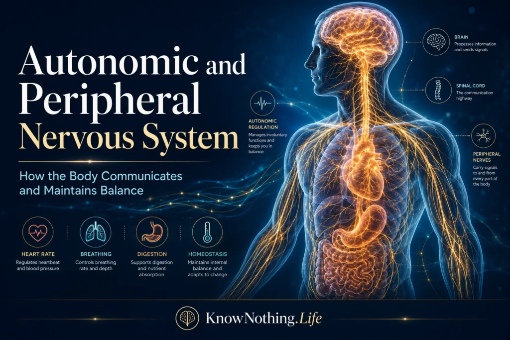
The brain and spinal cord could not guide the body without nerves extending into the skin, muscles, organs, blood vessels, and glands. This peripheral nervous system, or PNS, is the interface between the central nervous system and the body. It carries sensory information inward, sends motor commands outward, and regulates internal processes that rarely enter awareness. Anatomically and functionally, the PNS includes somatic sensory and motor pathways as well as autonomic pathways serving smooth muscle, cardiac muscle, and glands. Modern reviews describe the autonomic nervous system not merely as an emergency mechanism, but as a continuously active regulator of circulation, respiration, digestion, metabolism, and temperature.
Peripheral nerves are therefore more than passive cables. Receptors throughout the body detect pressure, temperature, tissue injury, chemical changes, stretch, and the condition of internal organs. Their signals enter the spinal cord and brain, where they are integrated with emotion, attention, memory, and current goals. Eduardo Benarroch’s influential 1993 review, “The Central Autonomic Network: Functional Organization, Dysfunction, and Perspective,” linked cortical, limbic, hypothalamic, and brainstem regions to visceral control. Later neuroimaging research identified the insula, amygdala, and midcingulate cortex in autonomic processing, linking bodily regulation to pain, emotion, motivation, and conscious experience.
Structure and Signaling in Peripheral Nerves
The PNS is commonly divided into sensory, motor, and autonomic components. Sensory, or afferent, fibers carry information from receptors toward the brain and spinal cord, while motor, or efferent, fibers carry commands to muscles and other targets. The somatic motor system usually employs a single neuron extending from the central nervous system to skeletal muscle. Autonomic motor pathways generally use a two-neuron chain: a preganglionic neuron communicates with a postganglionic neuron located in a peripheral ganglion. This arrangement helps distribute and coordinate signals before they reach organs.
Peripheral nerves contain axons of different diameters and conduction speeds, supported by connective tissue and Schwann cells. Myelin allows some axons to conduct impulses rapidly between nodes of Ranvier. Alan Hodgkin and Andrew Huxley’s landmark paper, “A Quantitative Description of Membrane Current and Its Application to Conduction and Excitation in Nerve,” established the ionic basis of the action potential, while later experiments demonstrated saltatory conduction in peripheral myelinated fibers. Schwann cells form peripheral myelin, organize nodal structures, support axonal metabolism, and respond to injury. Their roles help explain why peripheral axons sometimes regenerate better than central pathways, although recovery depends on injury severity and location.
Sympathetic, Parasympathetic, and Enteric Divisions
The autonomic nervous system is conventionally divided into sympathetic, parasympathetic, and enteric divisions. Sympathetic activity helps meet demands by increasing cardiac output, mobilizing energy, redirecting blood flow, widening the pupils, and supporting temperature regulation. Parasympathetic activity contributes to digestion, glandular secretion, energy conservation, and slowing the heart. These divisions are often described as “fight or flight” and “rest and digest,” but those phrases oversimplify a dynamic system. Both branches remain active at baseline, and their effects are not always direct opposites. Autonomic output shifts with posture, exercise, sleep, digestion, emotion, temperature, and disease.
The enteric nervous system consists of extensive neural circuits embedded within the gastrointestinal tract. It organizes local reflexes controlling motility, secretion, blood flow, and responses to intestinal contents while communicating with sympathetic and parasympathetic pathways. The vagus nerve carries sensory information toward the brain and motor signals back toward thoracic and abdominal organs. Research increasingly presents the vagus as part of a bidirectional monitoring system rather than simply a “calming nerve.” Vagal sensory fibers convey information about stretch, nutrients, inflammation, and organ state, helping central circuits adjust digestion, metabolism, circulation, and behavior.
Homeostasis, Reflexes, and Stress
Autonomic regulation is central to homeostasis, the preservation of viable internal conditions despite constant change. The arterial baroreflex provides a clear example. Stretch-sensitive receptors in major arteries detect blood-pressure changes and send information to the brainstem, which alters sympathetic and parasympathetic output to the heart and blood vessels. During standing, exercise, dehydration, or danger, this reflex supports rapid cardiovascular adjustment. Contemporary research shows that the baroreflex is not merely a rigid return-to-normal mechanism; it is continually reset according to behavior and physiological context. Heart rate variability is often used as a noninvasive indicator of autonomic regulation, although it is not a complete measure of sympathetic-parasympathetic “balance.”
Stress responses further demonstrate how autonomic function joins the brain, body, and environment. The insula, anterior cingulate cortex, amygdala, hypothalamus, periaqueductal gray, locus coeruleus, and brainstem nuclei help coordinate cardiovascular changes, vigilance, respiration, hormonal output, and defensive behavior. Acute sympathetic activation can be highly adaptive because it reallocates resources toward an immediate challenge. Persistent dysregulation, however, is associated with cardiovascular strain and disturbances in metabolism, sleep, and immune signaling. Early theories of emergency physiology established the importance of coordinated bodily responses, but modern neuroscience describes stress as a flexible pattern across interacting autonomic, endocrine, immune, and behavioral systems rather than one uniform reaction.
Sensation, Movement, Injury, and Disease
Somatic peripheral pathways make perception and voluntary movement possible. Sensory receptors convert physical and chemical events into neural signals representing touch, vibration, limb position, temperature, and pain. Motor axons activate skeletal muscles, while sensory feedback continually corrects movement as it unfolds. This loop supports posture, balance, dexterity, and coordination. Damage to large sensory fibers may impair vibration and position sense, whereas injury to small fibers may produce burning pain, abnormal temperature sensation, altered sweating, or other autonomic symptoms. Because routine nerve-conduction studies mainly assess large fibers, normal results do not exclude small-fiber neuropathy.
After severe peripheral axon injury, the separated distal segment undergoes Wallerian degeneration. Schwann cells and immune cells remove damaged material, and Schwann cells adopt a repair-supporting state that helps guide new growth. Degeneration is not simply decay; it creates conditions for regeneration. Yet axons must cross the injury site, enter the correct pathways, and reach their former targets before muscles and receptors deteriorate. Peripheral and autonomic disorders can result from diabetes, autoimmunity, infection, toxins, inherited mutations, trauma, nutritional deficiency, or neurodegeneration. Symptoms may include numbness, weakness, pain, orthostatic hypotension, abnormal sweating, constipation, bladder problems, or unstable heart rate.
Diagnosis, Treatment, and the Future of Neuromodulation
Clinical evaluation combines the symptom pattern, neurological examination, laboratory investigation, and tests selected for the fibers likely to be affected. Nerve-conduction studies and electromyography can reveal large-fiber and motor abnormalities. Blood-pressure and heart-rate responses during posture changes can expose cardiovascular autonomic failure, while sweat testing evaluates sympathetic pathways. When small-fiber neuropathy is suspected, skin biopsy can measure intraepidermal nerve fiber density and provide evidence missed by standard electrodiagnostic tests. This reflects a central lesson of peripheral neurology: similar symptoms may arise from damage to different sensory, motor, or autonomic pathways.
Peripheral nerves are also becoming therapeutic access points. In the landmark 2000 study “Vagus Nerve Stimulation Attenuates the Systemic Inflammatory Response to Endotoxin,” Linda Borovikova and colleagues found that vagus nerve stimulation reduced inflammatory signaling in an experimental model, helping establish the modern concept of neural regulation of immunity. Vagus nerve stimulation is used clinically for epilepsy, while researchers investigate neuromodulation for depression, inflammatory disease, gastrointestinal disorders, and metabolic conditions. Other strategies seek to improve peripheral nerve repair through engineered conduits, growth factors, electrical stimulation, and Schwann-cell-based therapies. These developments show that autonomic and peripheral systems are not secondary extensions of the brain. They are distributed systems of sensation, action, regulation, and repair through which the organism maintains itself and responds to the world.


