
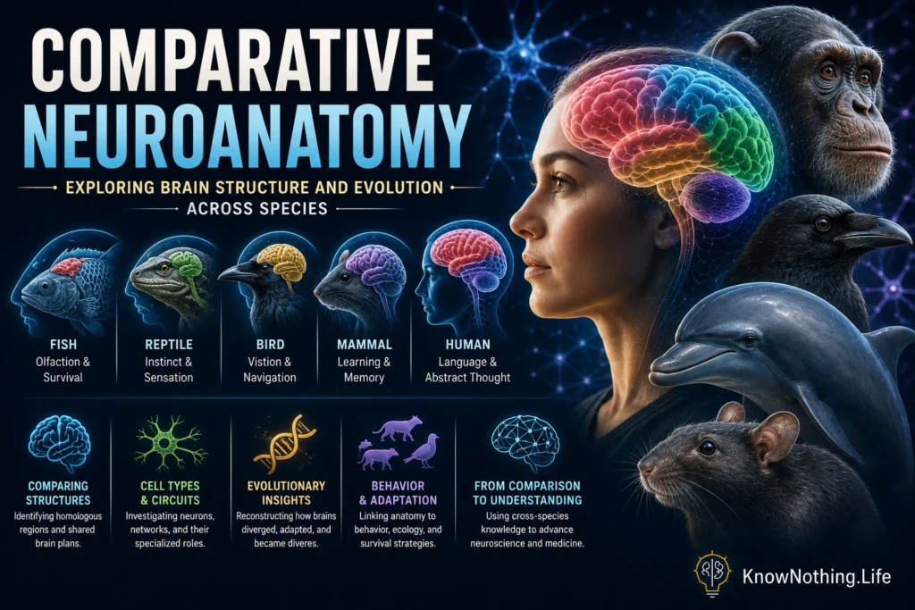
Comparative neuroanatomy examines the similarities and differences among nervous systems across species. By comparing the brains of mammals, birds, reptiles, amphibians, fish, insects, mollusks, and other animals, researchers can reconstruct how neural structures evolved and how anatomy supports particular behaviors. The field considers brain-region size, cellular composition, developmental origin, connectivity, gene expression, and microscopic organization. Its purpose is not to rank animals from primitive to advanced, but to identify inherited neural plans and the specialized modifications that emerged in different evolutionary lineages.
Every living nervous system represents a biological solution to a particular set of challenges. An owl requires neural systems that support precise hearing and vision, a bat must process returning echoes, and a mole depends heavily on touch and smell. Humans have extensive association cortices and prolonged neural development, but those characteristics are not universal standards by which other brains should be measured. Comparative neuroanatomy instead asks how each nervous system is adapted to an animal’s body, environment, behavior, and evolutionary history.
Homology, Analogy, and the Problem of Comparison
One of the field’s central challenges is determining whether structures in different species are homologous. Homologous structures are inherited from a common ancestral structure, even when they now differ in appearance or function. Analogous structures perform similar functions but evolved independently. Brain regions cannot be declared homologous merely because they occupy similar locations or participate in comparable behaviors. Researchers examine embryonic development, gene-expression patterns, cell types, connectivity, and relationships with surrounding structures to establish probable evolutionary correspondence.
The vertebrate pallium provides a famous example. The mammalian neocortex has a layered organization, while the corresponding forebrain regions of birds are arranged largely into clusters of cells rather than the same six-layered pattern. Earlier terminology treated much of the avian forebrain as an enlarged basal ganglia, reinforcing the mistaken impression that birds lacked cortical equivalents. Developmental, molecular, and connectivity evidence instead showed that substantial portions of the avian pallium share ancestry and functional characteristics with components of the mammalian pallium. The remaining debate concerns precisely how these regions should be matched across lineages and how often similar neural organizations evolved independently.
Shared Developmental Patterns and Mosaic Evolution
Brain regions often change together because their growth is linked by development. In a landmark 1995 study, Barbara Finlay and Richard Darlington analyzed the sizes of multiple brain structures across mammalian species. They found strong scaling relationships among regions and observed that structures produced later during development, particularly parts of the forebrain, tend to expand disproportionately as total brain size increases. Their results suggested that evolutionary alterations in developmental timing can enlarge several structures in coordinated ways rather than requiring completely separate modifications for every region.
Coordinated growth does not mean that the brain evolves only as a single unit. Robert Barton and Paul Harvey demonstrated in 2000 that mammalian brain regions also undergo mosaic evolution. They found that the relative size of the neocortex differed substantially between primates and insectivores after overall brain size was considered. Structures with close anatomical and functional relationships also tended to evolve together somewhat independently of other systems. Comparative neuroanatomy therefore combines two principles: developmental programs constrain how structures can change, while natural selection can selectively modify functionally connected systems.
Comparing Brain Size and Neuron Number
Brain size is one of the easiest anatomical measurements to collect, but it can also be misleading. Larger animals often require larger brains simply to control more muscles, process larger sensory surfaces, and regulate more internal tissue. Researchers therefore compare relative brain size, regional proportions, neuron numbers, cell density, and connectivity rather than relying on mass alone. Even these measurements must be interpreted carefully because the same volume of neural tissue can contain very different numbers and arrangements of neurons in different animal groups.
Research using the isotropic fractionator, a method that estimates cell numbers from suspended cell nuclei, transformed these comparisons. Suzana Herculano-Houzel and colleagues found that primate brains follow different neuronal scaling rules from rodent brains. As primate brains increase in size, neuron density declines less sharply, allowing a primate brain to contain more neurons than a similarly sized rodent brain. The human brain broadly follows this primate pattern, with its unusual capacities partly associated with the large absolute number of neurons contained in its cerebral cortex rather than with a completely unique anatomical plan.
Different Anatomies Can Support Complex Cognition
Bird brains reveal why comparative anatomy must look beyond superficial appearance. Birds lack a mammalian-style neocortex, yet parrots and corvids perform sophisticated tasks involving memory, flexible problem-solving, tool use, planning, and social learning. In 2016, Seweryn Olkowicz and colleagues counted neurons in the brains of 28 bird species. They found that parrots and songbirds pack exceptionally large numbers of neurons into their pallial regions, sometimes matching or exceeding the forebrain neuron counts of primates with much larger brains. Compact size does not necessarily indicate limited processing capacity.
Cephalopods provide an even more distant comparison. Octopuses evolved complex nervous systems independently from vertebrates, and much of their neural tissue is distributed through the arms rather than concentrated entirely in the central brain. Their anatomy supports flexible movement, tactile exploration, camouflage, and problem-solving through a partly decentralized system. Similar behavioral complexity can therefore emerge from radically different neural organizations. Comparative neuroanatomy identifies these cases of convergence while also showing that intelligence is not one ability represented by one structure.
Comparative Cellular Neuroanatomy
Modern neuroanatomy increasingly compares brains at the level of individual cell types. Traditional methods classified neurons by shape, location, neurotransmitter, and projection pattern. Single-cell and single-nucleus sequencing now allow researchers to compare thousands of cells according to the genes they express and the regulatory states of their DNA. These approaches can reveal whether apparently similar cells in two species share a conserved molecular identity or have diverged substantially during evolution.
Trygve Bakken and colleagues analyzed more than 450,000 nuclei from the primary motor cortices of humans, marmosets, macaques, and mice. Their 2021 study found broadly conserved neuronal and non-neuronal classes whose similarity reflected evolutionary distance. It also found species differences in cell proportions, gene expression, DNA methylation, chromatin accessibility, and specialized projection neurons. The results showed that homologous cortical systems can retain a common cellular foundation while modifying the molecular details that regulate connectivity, signaling, and physiology.
Comparing Connections and Neural Networks
The function of a brain region depends not only on its cells but also on its connections. Comparative connectomics studies how neural networks are organized across species using tract tracing, electron microscopy, diffusion imaging, functional imaging, and graph theory. Across many nervous systems, researchers find recurring architectural features. Connections tend to form modules that support specialized functions, while hubs and highly interconnected cores help information move between modules. These patterns reflect a compromise between efficient communication and the biological cost of building and maintaining long connections.
Comparisons among primates indicate extensive conservation in cortical and subcortical connectivity, but also important specialization. Humans and other primates share many major white-matter pathways and resting-state networks. Differences are especially relevant in regions and pathways involved in language, imitation, social cognition, and tool use, although imaging methods can produce false connections or miss real ones. Comparative connectomics must therefore establish anatomical homologies carefully and validate noninvasive findings against histological evidence whenever possible.
Why Model Organisms Are Both Valuable and Limited
Comparative neuroanatomy explains why animals can serve as models for human neuroscience. Conserved structures, neurotransmitters, developmental pathways, and cell types allow research in flies, fish, rodents, and nonhuman primates to reveal general principles of neural function. Studies of the mouse hippocampus, for example, can illuminate mechanisms of memory, while fruit-fly circuits can reveal fundamental rules of learning and sensory processing. Evolutionary continuity makes this translation possible.
Differences among species also set strict limits. A cell type may express different genes in a human and a mouse, a pathway may connect to different targets, or a cortical region may be expanded and reorganized in one lineage. Bakken and colleagues found both conserved motor-cortex cell classes and substantial species-specific molecular features, demonstrating why no model organism reproduces every aspect of human neural biology. Model selection should depend on the structure, disease, or behavior being studied rather than on convenience alone.
The Future of Comparative Neuroanatomy
New technologies are producing increasingly detailed atlases of species that were previously represented by only a few anatomical measurements. Spatial transcriptomics can map gene activity while preserving each cell’s position in the tissue. Three-dimensional imaging can trace long neuronal projections, and electron microscopy can reconstruct complete local circuits. In 2024, researchers created spatial single-cell atlases of the mouse, marmoset, and macaque cerebellum, identifying deeply conserved organization alongside primate-specific cell subtypes. Such projects connect gross anatomy with cellular identity and gene regulation.
The greatest value of comparative neuroanatomy is that it replaces simplistic rankings with an evolutionary explanation of neural diversity. Brains contain ancient structures, developmentally linked systems, specialized regions, conserved cell classes, and independently evolved solutions to similar problems. Human brains are distinctive in important respects, but they remain variations on biological themes shared across deep evolutionary time. By studying many species rather than treating one brain as the universal model, comparative neuroanatomy reveals both the common principles of nervous-system organization and the remarkable range of forms those principles can produce.


