
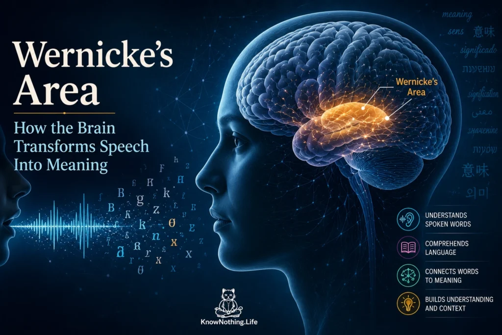
Wernicke’s area is a historically important language region associated with understanding spoken words and organizing meaningful speech. It is usually placed in the dominant cerebral hemisphere—most often the left—near the posterior superior temporal gyrus and adjacent temporal-parietal cortex. Traditional diagrams depict it as a clearly bounded “language-comprehension center,” but researchers have never agreed on one precise anatomical definition. Depending on the source, the label may include parts of the superior temporal gyrus, superior temporal sulcus, middle temporal gyrus, supramarginal gyrus, or angular gyrus. Modern research therefore treats Wernicke’s area as a variable portion of a larger language network rather than a single, universally located structure.
The region helps connect patterns of sound with linguistic representations, but it does not independently contain the meanings of every word or sentence. Speech comprehension requires the auditory system to analyze rapidly changing sounds, identify phonemes and words, retrieve semantic knowledge, interpret grammar, and integrate information over time. These operations are distributed across temporal, parietal, frontal, and subcortical systems connected through multiple white-matter pathways. Posterior temporal cortex remains important, but the familiar statement that Wernicke’s area “understands language” compresses several distinct processes into one misleading phrase.
Carl Wernicke and the Classical Language Model
German physician Carl Wernicke developed his influential theory after studying patients whose language difficulties differed from the nonfluent speech described earlier by Paul Broca. In his 1874 work, Der aphasische Symptomencomplex, Wernicke described people who could speak with normal rhythm and apparent ease but had difficulty understanding language and producing meaningful verbal content. He associated the syndrome with damage in the posterior portion of the left temporal lobe, near auditory cortex. His observations helped establish the idea that language contains partly separable components and that different forms of aphasia can follow damage to different parts of the brain.
Wernicke also proposed that language depended on communication between sensory and motor representations. In the classical model, posterior temporal cortex stored auditory word images, Broca’s area supported the motor patterns of speech, and a connecting pathway allowed heard words to influence verbal production. Damage to the connection could theoretically produce conduction aphasia, in which speech and comprehension remain relatively preserved but repetition becomes impaired. This network concept was remarkably forward-looking, although the anatomical details were incomplete. Contemporary neuroscience supports distributed pathways and functional interactions, but it no longer accepts that two cortical centers alone can explain language.
Where Is Wernicke’s Area?
The exact location of Wernicke’s area remains controversial partly because the term has been used to describe anatomy, function, and a clinical syndrome as though they were the same thing. Anatomically, it is often identified with the posterior superior temporal gyrus. Functionally, it may refer more broadly to tissue involved in recognizing speech or retrieving phonological forms. Clinically, it is sometimes defined as the region damaged in Wernicke’s aphasia. These definitions do not consistently point to identical tissue, and the cortical areas activated during comprehension vary across individuals and tasks.
Lesion research has especially challenged the textbook boundaries. Nina Dronkers and colleagues studied patients with chronic aphasia and found that comprehension deficits were associated with damage to several regions, including the posterior middle temporal gyrus, anterior superior temporal gyrus, superior temporal sulcus, angular gyrus, and parts of the frontal cortex. Damage restricted to the classically defined Wernicke’s area was not significantly associated with impaired comprehension on their principal measure. The findings did not make posterior temporal cortex irrelevant; they showed that language understanding cannot be localized by drawing one circle on the cortical surface.
From Sound to Recognizable Speech
Before words can be understood, the auditory system must separate speech from other sounds and represent features such as frequency, timing, pitch, and phonetic structure. Primary auditory cortex in Heschl’s gyrus responds rapidly to acoustic information, while surrounding nonprimary regions of the superior temporal cortex show greater sensitivity to complex speech patterns. Older models often proposed a serial hierarchy in which basic sound was processed first and then passed forward to a separate linguistic center. Direct recordings indicate that the system is more parallel and distributed than this account suggests.
In 2021, Liberty Hamilton, Adeen Flinker, and colleagues combined intracranial recordings, electrical stimulation, and evidence from surgical removal to compare primary and nonprimary auditory cortex. They found parallel processing across these regions rather than a simple step-by-step transformation from sound to language. Stimulating primary auditory cortex could produce auditory sensations without necessarily disrupting speech perception, whereas stimulation of nonprimary superior temporal regions could interfere with the perception of spoken language. The study supports an essential role for lateral superior temporal cortex in speech analysis while showing that this function is distributed across specialized auditory populations.
Words, Meaning, and Sentence Comprehension
Recognizing a spoken word requires more than accurately hearing its sounds. The listener must connect the auditory sequence to a familiar lexical representation and retrieve relevant conceptual knowledge. Research indicates that posterior temporal regions contribute strongly to phonological processing and the recognition of word forms, while semantic knowledge depends heavily on a wider temporal network that includes the anterior temporal lobes. Damage to posterior temporal cortex can disrupt the ability to identify or repeat words, but damage to anterior temporal regions may produce especially severe loss of word meaning.
Marsel Mesulam and colleagues examined patients with primary progressive aphasia, a group of neurodegenerative disorders that can selectively damage different parts of the language network. Their analysis of 72 patients found that word and sentence comprehension could be dissociated and that no single circumscribed cortical area appeared equally essential for both. Severe word-comprehension impairment was most closely associated with anterior temporal damage, whereas sentence comprehension depended more heavily on a broader left-hemisphere network. The results directly challenged the idea that the traditional Wernicke region is the brain’s central storehouse of linguistic meaning.
Sentence comprehension adds demands that single-word recognition does not. The brain must combine meanings, follow grammatical relationships, maintain information in memory, and revise interpretations when later words change the apparent structure. A lesion-mapping study led by S. B. Pillay found that difficulty understanding phrases and sentences was strongly associated with damage to the mid-to-posterior middle temporal gyrus. More recent work involving 131 stroke survivors implicated the left superior and middle temporal regions together with several underlying white-matter pathways. These findings suggest that posterior temporal cortex helps integrate lexical, semantic, and syntactic information across multiword expressions.
Wernicke’s Area and Speech Production
Wernicke’s area is usually discussed in relation to comprehension, but posterior temporal systems also contribute to speech production. Before a word can be spoken, its sound structure must be retrieved and arranged in the correct sequence. Damage in posterior temporal and inferior parietal regions can interfere with phonological retrieval, producing sound substitutions, invented words, and impaired repetition. The speaker may retain fluent articulation because frontal and motor systems are functioning, yet the words selected or assembled may not accurately express the intended message.
This helps explain why fluent speech in Wernicke’s aphasia can contain phonemic paraphasias, semantic substitutions, vague expressions, or neologisms. The difficulty is not merely that a person cannot understand what others say. Disrupted monitoring and phonological selection can also prevent the speaker from recognizing or correcting errors in their own output. Posterior temporoparietal cortex appears particularly important for repetition, which requires heard speech to be maintained and converted into a form that can guide articulation. Mesulam and colleagues found that damage in this region severely affected repetition even when basic word comprehension was supported elsewhere.
Wernicke’s Aphasia
Wernicke’s aphasia, sometimes called fluent or receptive aphasia, most commonly follows a stroke affecting the posterior part of the left cerebral hemisphere. Speech may remain effortless, melodic, and grammatically shaped, but its content can be difficult to follow. People may substitute incorrect words, combine phrases without a coherent message, invent word-like forms, or speak at great length without conveying the intended information. Comprehension and repetition are usually impaired, while reading and writing may show related disturbances. The exact symptom pattern varies because lesions differ in size and frequently extend beyond the cortical territory traditionally labeled Wernicke’s area.
Calling the condition “receptive aphasia” can be misleading because language production is also disturbed. Likewise, fluent speech does not mean that speech is normal. Fluency describes rate, phrase length, melody, and articulatory ease, not accuracy or communicative effectiveness. Some patients are initially unaware of the extent of their errors, although awareness may improve as recovery progresses. Clinical evaluation therefore examines auditory comprehension, naming, repetition, reading, writing, discourse, semantic knowledge, and the ability to monitor communication rather than relying on fluency alone.
Connections, White Matter, and the Language Network
Posterior temporal cortex communicates with frontal and parietal regions through several dorsal and ventral pathways. Dorsal pathways help connect auditory and phonological representations with frontal speech-planning systems, while ventral pathways contribute to mapping words and sentences onto meaning. A stroke near Wernicke’s area may damage several tracts simultaneously, disconnecting distant areas whose cortical tissue remains intact. The resulting symptoms may therefore arise from interruption of a network rather than destruction of one comprehension center.
William Matchin and colleagues used connectome-based lesion-symptom mapping to reconsider the “Wernicke conundrum”: why lesions near the traditional area are associated with severe comprehension problems even though damage limited to some of its cortical portions may not be sufficient. Their results supported the importance of posterior temporal regions and associated connections in word and sentence comprehension. The study illustrates how cortical damage and disconnection can work together, producing broader deficits than would be predicted from the visible surface lesion alone.
Recovery and the Modern Interpretation
Recovery from Wernicke’s aphasia depends on lesion location, lesion size, the condition of surviving language pathways, age, overall health, and access to rehabilitation. Speech-language therapy may target comprehension, word retrieval, monitoring, reading, writing, and conversational repair. Improvement generally reflects the reorganization of surviving portions of the left-hemisphere network, supported in some cases by right-hemisphere regions and domain-general systems. A large longitudinal study of aphasia during the first year after stroke found that the location and extent of damage—especially within posterior perisylvian regions—were major determinants of outcome.
The modern interpretation preserves Wernicke’s fundamental insight that posterior temporal cortex is crucial to language while rejecting the idea of a single comprehension center. The region contributes to speech perception, phonological representation, word retrieval, repetition, and the integration of information across sentences. Meaning itself depends on broader temporal and frontal networks, and severe aphasia often reflects damage to both cortical areas and their connections. Wernicke’s area remains one of neuroscience’s most important landmarks, not because the nineteenth-century model was complete, but because it opened the way to understanding language as an organized system of interacting brain regions.


