
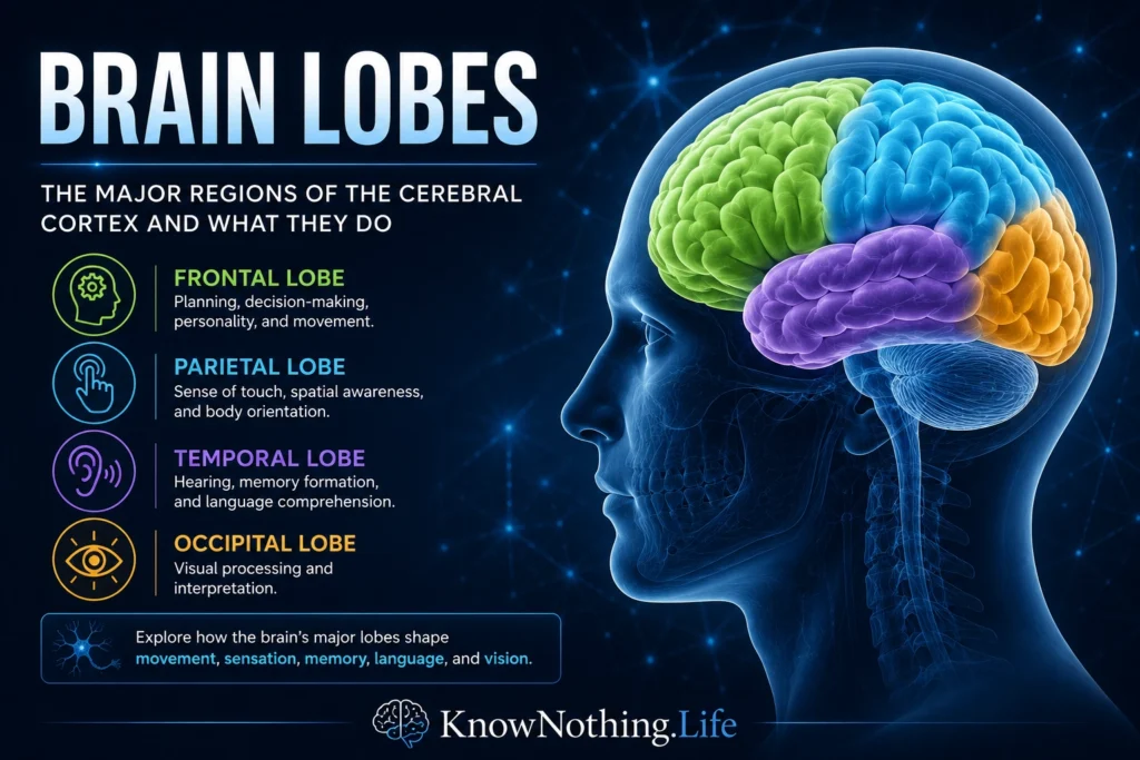
Brain lobes are the major anatomical regions of the cerebral cortex, the folded outer layer of the cerebrum involved in perception, voluntary movement, language, memory, reasoning, emotion, and conscious awareness. The four classical lobes are the frontal, parietal, temporal, and occipital lobes. These divisions are partly based on visible landmarks such as grooves, folds, and borders of the skull, but they also reflect important functional differences.
The lobes should not be understood as isolated boxes with one job each. A person reading a sentence, recognizing a face, reaching for a cup, or making a moral decision is not using one lobe alone. Brain function is distributed across networks that cross lobe boundaries. Still, the lobe system remains useful because it gives us a map of where major functions tend to cluster. The frontal lobe is strongly associated with planning and voluntary action; the parietal lobe with sensation and spatial awareness; the temporal lobe with hearing, memory, and meaning; and the occipital lobe with vision. Together, these lobes help turn raw sensation into perception, movement into skilled action, and experience into thought.
The Frontal Lobe
The frontal lobe sits at the front of the brain, behind the forehead, and is often associated with the most visibly “executive” aspects of human behavior. It includes the primary motor cortex, which helps control voluntary movement, as well as premotor and prefrontal regions involved in planning, attention, working memory, impulse control, social judgment, decision-making, and personality. The frontal lobe does not simply make people rational; it helps organize behavior toward goals. When someone resists an impulse, chooses words carefully, plans for the future, or adjusts behavior to fit a social situation, frontal-lobe networks are heavily involved.
The history of neuroscience often uses the famous case of Phineas Gage to illustrate the importance of frontal regions. Gage survived an 1848 accident in which an iron rod passed through his skull, damaging frontal areas. Later accounts of his personality change have sometimes been exaggerated, but the case remains influential because it helped connect frontal-lobe injury with changes in judgment, emotion, and social behavior. The frontal lobe is also central to language production, especially in the left inferior frontal region historically associated with Paul Broca’s work on aphasia. Broca’s nineteenth-century studies showed that damage to specific frontal areas could severely impair speech production while leaving many other abilities partly intact, helping establish the idea that complex mental functions have biological organization.
The Parietal Lobe
The parietal lobe lies behind the frontal lobe and above the temporal lobe. Its most basic role involves bodily sensation. The primary somatosensory cortex, located in the postcentral gyrus, receives information about touch, pressure, pain, temperature, and body position. This is why parietal-lobe damage can affect a person’s ability to locate touch, recognize objects by feel, or understand where parts of the body are in space. But the parietal lobe is not merely a touch-processing center. It helps integrate sensory information into a practical model of the body and surrounding world.
This integrative role makes the parietal lobe essential for spatial awareness, attention, hand-eye coordination, and movement guidance. It helps answer questions the brain must solve constantly: Where is my body? Where is that object? How far away is it? How should I move to reach it? Damage to the right parietal lobe can produce hemispatial neglect, in which a person may ignore the left side of space even though the eyes themselves are functioning. This shows that perception is not just the arrival of sensory data. The brain must organize attention, space, and body awareness into a coherent scene. The parietal lobe is one of the major regions that makes this possible.
The Temporal Lobe
The temporal lobe sits on the side of the brain, roughly behind the temples and beneath the lateral fissure. It is deeply involved in hearing, language comprehension, memory, object recognition, and emotion. The primary auditory cortex receives sound information, while surrounding areas help interpret speech, music, tone, and environmental sounds. The temporal lobe also contains medial structures, including the hippocampal region, that are crucial for forming new declarative memories. The famous patient H.M., studied by William Scoville and Brenda Milner, showed how medial temporal-lobe damage could severely impair the ability to form new long-term memories while leaving other abilities partly preserved. Later reviews describe H.M. as central to the development of modern memory neuroscience.
Language comprehension is another major temporal-lobe function. Carl Wernicke’s nineteenth-century work helped identify a posterior temporal region associated with understanding spoken language. Modern research has complicated the old idea of a single “Wernicke’s area,” showing that language comprehension depends on broader networks rather than one isolated patch of cortex. Neuroscientist Jeffrey Binder’s review of Wernicke’s area argues that the traditional label has often been applied too loosely and that modern evidence supports a more distributed model of language comprehension. The larger lesson is important: the temporal lobe helps connect sound, memory, recognition, and meaning, but it does so as part of a networked brain.
The Occipital Lobe
The occipital lobe is located at the back of the brain and is the primary cortical region for vision. Visual information travels from the eyes through the optic nerves and thalamus before reaching the primary visual cortex in the occipital lobe. From there, visual processing spreads into additional regions that help identify color, form, motion, depth, and object meaning. Seeing is not simply a camera-like recording of the world. The brain actively constructs visual experience by detecting edges, contrast, movement, spatial relationships, and patterns.
The work of David Hubel and Torsten Wiesel transformed the scientific understanding of visual cortex. Their studies showed that neurons in visual cortex respond selectively to specific features such as line orientation, helping reveal how the brain breaks visual information into organized components. Hubel shared the 1981 Nobel Prize in Physiology or Medicine for discoveries concerning information processing in the visual system, including the functional architecture of the visual cortex. The occipital lobe therefore demonstrates one of the brain’s central principles: perception is an active biological process. The world appears seamless, but the brain builds that seamless experience through specialized processing.
The Limbic Lobe, Insula, and Modern Brain Mapping
Although the four-lobe model is the most familiar, many neuroscientists also discuss the limbic lobe and the insula. The limbic lobe includes cortical and nearby structures involved in emotion, motivation, memory, and survival-related behavior. It overlaps with regions such as the cingulate cortex, parahippocampal areas, hippocampus, and amygdala-related networks. The insula lies deep within the lateral sulcus, hidden beneath parts of the frontal, parietal, and temporal lobes. It is involved in interoception, taste, pain, emotion, self-awareness, and the sense of the body’s internal state. These additions remind us that the classical lobe map is useful but incomplete.
Modern brain mapping also goes beyond surface anatomy. In 1909, Korbinian Brodmann divided the cerebral cortex into areas based on cytoarchitecture, meaning differences in cellular organization. His map remains influential, but later researchers have refined it using histology, neuroimaging, connectivity studies, and functional analysis. A review by Karl Zilles and colleagues notes that Brodmann’s maps still dominate his legacy, even though modern work has expanded our understanding of cortical organization. This matters because brain lobes are only one level of organization. Beneath the lobe labels are smaller cortical areas, networks, cell types, pathways, and dynamic patterns of activity.
Why Brain Lobes Matter
Brain lobes matter because they provide a practical map of the mind’s biological foundation. They help explain why different injuries cause different symptoms, why a stroke in one area may affect speech while another affects vision, and why memory, movement, attention, and perception depend on different but connected regions. The lobe system also gives students and general readers a way to begin understanding brain anatomy without getting lost in thousands of specialized terms. It is not the whole story, but it is a useful first map.
At the same time, the brain’s real power comes from coordination across lobes. The frontal lobe may plan a movement, the parietal lobe may guide it through space, the occipital lobe may supply visual information, and the temporal lobe may connect the action to memory and meaning. Human experience is not produced by one lobe at a time. It emerges from networks that integrate sensation, movement, attention, memory, language, emotion, and self-awareness. To study the brain lobes is to study both specialization and unity: the way different regions do different things, and the way the whole brain turns those differences into a single living mind.


