
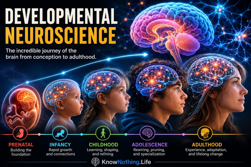
Developmental neuroscience is the scientific study of how the nervous system forms, changes, and becomes capable of producing perception, movement, emotion, learning, language, and social behavior. It examines development from the earliest stages of embryonic life through infancy, childhood, adolescence, and adulthood. Researchers investigate how genes guide the production of neural cells, how neurons reach their proper locations, how brain circuits become connected, and how experience modifies those circuits over time. The field combines neuroscience with embryology, genetics, psychology, medicine, education, and the study of human development.
Brain development is not a simple process in which structures appear one after another according to a fixed genetic blueprint. Genes provide essential instructions, but neural development also depends on chemical signals, spontaneous activity, sensory input, nutrition, hormones, relationships, and learning. Some developmental processes occur within relatively narrow periods, while others continue for decades. The mature brain is therefore the product of continuous interaction between biological preparation and experience.
Building the Brain Before Birth
The nervous system begins as a sheet of embryonic tissue that folds to form the neural tube, the structure from which the brain and spinal cord develop. Neural stem and progenitor cells then divide rapidly, producing neurons and glial cells. Newly generated neurons must travel from their places of origin to their eventual locations, acquire specialized identities, extend axons and dendrites, and establish connections with appropriate targets. These events require precisely timed interactions among genes, molecular signals, neighboring cells, and electrical activity.
Pasko Rakic’s research on cortical development helped explain how neurons become organized into the layered cerebral cortex. His radial unit hypothesis proposed that groups of neurons generated from progenitor cells near the cerebral ventricles migrate outward along radial glial fibers, forming vertical units that contribute to the organization and expansion of the cortex. Rakic’s work also demonstrated that neuronal migration is an active, guided process rather than the passive displacement of cells. Disruptions to proliferation, migration, or positioning can alter later circuit formation and contribute to cortical malformations and neurodevelopmental difficulties.
Prenatal development is both highly organized and vulnerable. Alcohol, certain infections, nutritional deficiencies, toxic exposures, and disruptions in placental function can affect neural growth, although outcomes depend on timing, intensity, genetic susceptibility, and other conditions. Recent longitudinal imaging research has continued to find associations between prenatal alcohol exposure and altered development of cortical and white-matter systems during childhood. Such findings do not imply that every exposure produces the same outcome, but they show that events occurring before birth can influence neural trajectories for years afterward.
Synapse Formation, Pruning, and Myelination
After neurons reach their destinations, they form synapses through which they communicate. Early development involves an enormous increase in synaptic connections, but the brain does not retain every connection it produces. Peter Huttenlocher’s postmortem studies of the human cortex showed that synaptic density rises rapidly during early life and later declines toward adult levels. His later work with Arun Dabholkar found that the timing of synapse formation and elimination differs across cortical regions, with sensory areas generally maturing earlier than association regions involved in more complex cognition.
This reduction is commonly called synaptic pruning, although the process is more selective than the term may suggest. Frequently used and well-coordinated connections are more likely to be stabilized, while other connections weaken or disappear. Pruning can make networks more efficient, but development is not simply a movement from “more synapses” to “fewer synapses.” Dendrites grow, axons change, neurotransmitter systems mature, and patterns of communication are continuously reorganized.
Myelination is another essential process. Myelin is a fatty insulating material formed around many axons, allowing signals to travel more rapidly and reliably. Longitudinal imaging studies show that white-matter organization continues to change throughout childhood and adolescence, with different pathways following different developmental schedules. Christian Beaulieu, Catherine Lebel, and colleagues found that the structural development of human brain wiring continues well beyond early childhood, while Jay Giedd and colleagues documented prolonged changes in major structures and white matter during childhood and adolescence.
Critical Periods and Experience-Dependent Plasticity
Some neural systems are especially sensitive to experience during particular stages of development. A critical period is a limited interval during which specific experience is necessary for a function to develop normally. A sensitive period is a broader interval during which experience has an unusually strong effect, even though learning may remain possible later. These concepts emerged partly from research on the developing visual system.
David Hubel and Torsten Wiesel showed that closing one eye during an early period of a kitten’s development profoundly altered responses in the visual cortex. Neurons that would normally respond to both eyes became dominated by the open eye, and cells in pathways serving the deprived eye showed physiological and anatomical changes. The same deprivation had much weaker effects when it occurred after the sensitive developmental window. Their work demonstrated that neural organization depends not only on inherited structure but also on patterned experience arriving at the right time.
Experience-dependent plasticity extends beyond vision. Exposure to language, movement, music, social interaction, and education changes the circuits used to process those experiences. Plasticity does not mean that the young brain can recover perfectly from every disruption or that development becomes fixed after childhood. Instead, the form and degree of plasticity change with age. Early brains may reorganize extensively, while mature brains retain more established networks but remain capable of learning, adaptation, and partial recovery after injury.
Caregiving, Stress, and the Developing Brain
Early relationships provide much of the stimulation through which infants learn to regulate attention, emotion, and stress. Responsive caregivers do more than satisfy physical needs. They help organize sleep, arousal, communication, exploration, and the interpretation of unfamiliar events. When caregiving is severely limited or unpredictable, development may be affected across cognitive, emotional, and neural systems.
The Bucharest Early Intervention Project provided rare experimental evidence about the effects of early psychosocial deprivation. Children who had been raised in Romanian institutions were randomly assigned either to continued institutional care or to specially developed foster care. Those placed in foster families showed better cognitive outcomes than children who remained in institutional care, particularly when placement occurred earlier. Follow-up research found differences in brain electrical activity and white-matter development, while stable, high-quality caregiving was associated with more typical developmental patterns.
Stress is not inherently damaging; manageable challenges can support learning and adaptation. Persistent or extreme stress, however, may repeatedly activate hormonal and autonomic systems while the brain is still developing. Research has linked prenatal and early-life stress with differences in the development and connectivity of the amygdala, hippocampus, and prefrontal cortex, although effects vary according to timing, severity, sex, later caregiving, and other influences. These findings describe altered probabilities rather than inevitable outcomes. Supportive relationships can moderate risk and help children develop more effective emotional regulation.
Childhood and Adolescent Brain Development
Brain development continues throughout childhood as sensory, motor, language, memory, and executive systems become more coordinated. Longitudinal magnetic resonance imaging has shown that cortical development follows a complex regional sequence rather than occurring uniformly across the brain. In a study mapping participants from ages four to twenty-one, Nitin Gogtay and colleagues found that changes generally progressed from regions supporting basic sensory and motor functions toward association areas involved in planning, attention, and higher-order cognition.
Adolescence is not simply the final stage of childhood development. It is a period of extensive biological and social transition in which reward sensitivity, emotional significance, peer relationships, and opportunities for independent decision-making change rapidly. B. J. Casey, Rebecca Jones, and Todd Hare proposed that adolescent behavior reflects an imbalance in the developmental timing of neural systems: reward and emotional circuits may become highly responsive while prefrontal systems supporting regulation and long-term planning are still maturing. This model does not portray adolescents as incapable of rational thought. It suggests that behavior becomes especially sensitive to emotion, social context, and immediate incentives under certain conditions.
The prolonged development of the prefrontal cortex and its connections supports improvements in working memory, inhibition, planning, and flexible decision-making. At the same time, adolescence creates opportunities for exploration, learning, creativity, and adaptation. Increased sensitivity to social and rewarding experiences can encourage risky behavior, but it can also support education, friendship, identity formation, and the acquisition of adult skills.
Developmental Disorders and Individual Differences
Developmental neuroscience contributes to the study of autism, attention-deficit/hyperactivity disorder, intellectual disability, epilepsy, dyslexia, cerebral palsy, and other conditions affecting cognition or behavior. These conditions cannot usually be reduced to one damaged location. They may involve differences in cellular development, synaptic signaling, network connectivity, timing, or the coordination of several systems. The same diagnosis can also involve different biological pathways in different individuals.
Developmental findings must be interpreted carefully. A difference in brain structure or activation is not automatically a deficit, and group averages cannot determine the abilities or future of one child. Developmental pathways are influenced by genetics, health, education, culture, relationships, and opportunity. Neural diversity is expected, and many forms of variation are compatible with healthy and meaningful lives.
Developmental neuroscience ultimately reveals a brain that is neither completely predetermined nor infinitely malleable. Neural development follows biological constraints, but experience helps decide which connections are strengthened, which skills become practiced, and how individuals adapt to their surroundings. From prenatal cell migration to adolescent decision-making, the developing brain is continually constructing itself through an exchange among genes, bodies, relationships, and environments.


