
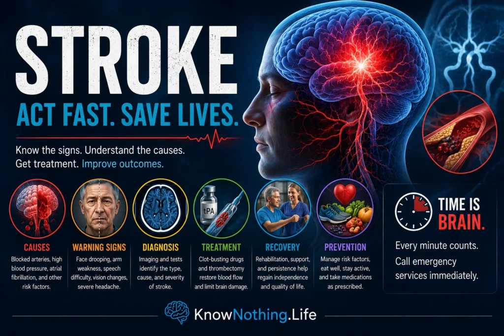
A stroke occurs when part of the brain suddenly loses its normal blood supply or when a damaged blood vessel releases blood into or around brain tissue. Without adequate circulation, neurons are deprived of oxygen and glucose, disrupting the electrical and chemical processes required for movement, language, memory, sensation, and consciousness. Brain cells can become injured within minutes, which is why stroke is treated as an immediate medical emergency rather than a condition that can be observed at home. Depending on the location and severity of the injury, a stroke may cause temporary impairment, permanent disability, or death.
Although stroke is often associated with older adults, it can occur at any age. Its effects vary because different brain regions perform different functions. Damage to motor pathways may produce weakness or paralysis, while injury to language networks may impair speaking or understanding. A stroke affecting the cerebellum or brainstem can disturb balance, coordination, swallowing, breathing, or consciousness. The clinical syndrome therefore depends not only on the amount of damaged tissue but also on which neural networks have been interrupted.
Ischemic and Hemorrhagic Stroke
Ischemic stroke is the most common form and develops when a clot, plaque, or other obstruction prevents blood from reaching part of the brain. Some clots form within arteries narrowed by atherosclerosis, while others travel from the heart or a more distant blood vessel. Atrial fibrillation is an especially important cardiac cause because irregular contractions can allow blood to pool and form clots that later enter the cerebral circulation. The affected region usually contains a severely damaged central core surrounded by an area of threatened but potentially salvageable tissue known as the ischemic penumbra.
Hemorrhagic stroke occurs when a vessel ruptures and blood accumulates within the brain or in the surrounding spaces. Intracerebral hemorrhage refers to bleeding directly into brain tissue, while subarachnoid hemorrhage commonly results from the rupture of an aneurysm into the space surrounding the brain. Injury results from the initial bleeding, pressure created by the expanding blood collection, inflammation, and disruption of nearby circulation. High blood pressure is an important cause of intracerebral hemorrhage because chronic pressure can weaken small arteries over time.
Transient Ischemic Attacks and Warning Signs
A transient ischemic attack, or TIA, produces stroke-like symptoms caused by a temporary interruption of blood flow without evidence of lasting brain infarction. The symptoms may disappear within minutes, but their resolution does not make the event harmless. A TIA can indicate unstable vascular disease, a cardiac source of embolism, or another condition capable of producing a larger stroke. Because it is impossible to determine immediately whether symptoms will resolve, suspected TIAs require the same urgent evaluation as completed strokes.
Common warning signs include sudden facial drooping, weakness or numbness affecting one side of the body, difficulty speaking, confusion, loss of vision, severe imbalance, or an unexplained intense headache. The acronym BE FAST adds balance and eye or vision problems to the familiar signs involving the face, arms, speech, and time. Symptoms may be subtle, particularly when the stroke affects vision, coordination, or cognition rather than producing obvious paralysis. Emergency services should be contacted immediately because treatment eligibility often depends on when the person was last known to be neurologically normal.
Causes and Preventable Risk Factors
Stroke develops through an interaction of age, genetics, vascular disease, cardiac health, behavior, and environmental conditions. High blood pressure is the most important modifiable risk factor because it accelerates atherosclerosis, damages small vessels, and increases the likelihood of both blocked and ruptured arteries. Other major contributors include smoking, diabetes, abnormal blood lipids, physical inactivity, obesity, unhealthy diet, excessive alcohol consumption, atrial fibrillation, and psychosocial stress.
The international INTERSTROKE study, led by Martin O’Donnell and colleagues, examined patients across diverse regions and populations. Its 2016 analysis concluded that ten potentially modifiable factors were collectively associated with roughly 90 percent of the population-attributable risk of stroke. The findings do not mean that every individual stroke is preventable, but they demonstrate that controlling blood pressure, avoiding tobacco, remaining physically active, treating metabolic disease, and addressing cardiac risks could substantially reduce the global burden.
Diagnosis and the Race to Restore Blood Flow
Emergency diagnosis begins with neurological assessment and brain imaging. Computed tomography can rapidly identify bleeding, while CT angiography can reveal an obstructed major artery. Magnetic resonance imaging is highly sensitive to early ischemic injury, and perfusion imaging can estimate how much tissue has already infarcted and how much might still be rescued. Blood tests, cardiac monitoring, vessel imaging, and later investigations help determine whether the stroke originated from atherosclerosis, small-vessel disease, atrial fibrillation, arterial dissection, a clotting disorder, or another cause.
The 1995 National Institute of Neurological Disorders and Stroke trial transformed acute care by showing that intravenous tissue plasminogen activator, now generally called alteplase, improved functional outcomes when administered within three hours of ischemic stroke onset. Treatment increased the risk of symptomatic brain hemorrhage, making rapid imaging and careful selection essential. Later research, including the ECASS III trial, supported treatment for selected patients within an extended window of up to four and a half hours. These studies established the principle that earlier restoration of circulation generally offers a greater chance of preserving brain function.
Mechanical Thrombectomy
Intravenous thrombolysis may not dissolve large clots blocking major cerebral arteries. Mechanical thrombectomy addresses this problem by guiding a catheter through the circulation and using a specialized device to remove the obstruction. Earlier endovascular trials produced disappointing results, partly because they used older technology and did not consistently confirm large-vessel occlusion before treatment. The development of stent retrievers, faster imaging, and improved patient selection changed the field.
The MR CLEAN trial, published in 2015 by Olvert Berkhemer and colleagues, demonstrated that intraarterial treatment improved outcomes for patients with proximal anterior-circulation occlusions treated within six hours. The DAWN and DEFUSE 3 trials later showed that selected patients could benefit six to 24 hours after they were last known to be well when imaging revealed a relatively small completed infarct and substantial salvageable tissue. These findings shifted treatment from a simple clock-based model toward a “tissue clock,” recognizing that the rate of brain injury differs among patients.
Treating Hemorrhagic Stroke
Hemorrhagic stroke requires a different strategy because clot-dissolving medication would worsen bleeding. Treatment may include controlling blood pressure, reversing anticoagulant drugs, reducing dangerous pressure inside the skull, managing seizures, and treating the underlying vascular abnormality. A ruptured aneurysm may be sealed through surgical clipping or endovascular coiling, while selected intracerebral hemorrhages may require surgical evacuation. Management depends on the bleeding location, volume, rate of expansion, neurological condition, and general health of the patient.
The INTERACT2 trial, led by Craig Anderson and colleagues, investigated intensive blood-pressure reduction following spontaneous intracerebral hemorrhage. Intensive treatment did not significantly reduce the trial’s primary outcome of death or major disability, although analysis across the full disability scale suggested improved functional outcomes. The later ATACH-2 trial found that lowering systolic pressure to a more aggressive target did not provide additional benefit and increased renal adverse events. Together, these studies illustrate that blood pressure must be reduced carefully rather than as rapidly or extensively as possible.
Rehabilitation and Neural Recovery
Surviving the initial stroke is often the beginning of a longer recovery process. Rehabilitation may involve physical therapy for mobility and balance, occupational therapy for daily activities, speech-language therapy for communication and swallowing, and neuropsychological care for cognitive or emotional changes. Recovery reflects resolution of temporary dysfunction, compensation by surviving networks, relearning through practice, and neuroplastic changes in how the brain organizes movement and behavior. Improvement can continue long after hospitalization, although the speed and extent of recovery vary widely.
More therapy is not always beneficial when introduced at the wrong intensity or time. The AVERT trial examined very early, frequent mobilization beginning within 24 hours and found that this intensive approach did not improve outcomes compared with usual care. The result challenged the assumption that immediately increasing activity must always accelerate recovery. Modern rehabilitation is increasingly individualized according to medical stability, impairment, endurance, personal goals, and the type of function being retrained.
Preventing Another Stroke
After an ischemic stroke or TIA, prevention depends on identifying the cause. Antiplatelet drugs are commonly used for noncardioembolic stroke, while anticoagulants are generally more appropriate when atrial fibrillation is responsible. Treatment may also include blood-pressure control, cholesterol-lowering therapy, diabetes management, smoking cessation, improved diet, exercise, and procedures for severe carotid narrowing. In the CHANCE and POINT trials, short-term treatment with clopidogrel and aspirin reduced early recurrent ischemic events after certain minor strokes or high-risk TIAs, although prolonged dual therapy increased bleeding risk.
The future of stroke care will combine faster emergency systems with increasingly precise patient selection. Artificial-intelligence-assisted imaging, mobile stroke units, improved clot-removal devices, blood-based biomarkers, neuroprotective therapies, and brain-stimulation approaches may expand the number of people who benefit from treatment. Yet the greatest population-level opportunity remains prevention. Stroke is an acute brain emergency, but it is also frequently the final event in a vascular process that has developed silently for years. Effective care therefore begins before symptoms appear and continues through emergency treatment, rehabilitation, and lifelong risk reduction.


