
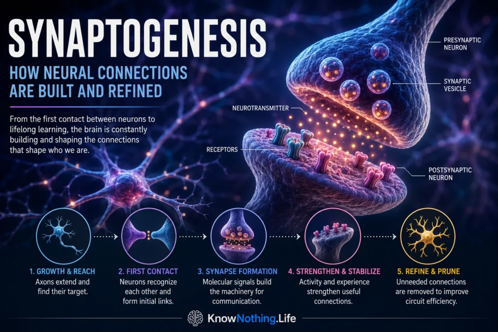
Synaptogenesis is the process through which neurons form specialized communication points called synapses. At a chemical synapse, the presynaptic neuron releases neurotransmitters from an axon terminal, while receptors and signaling proteins on the postsynaptic cell detect and respond to those chemicals. Most synapses in the central nervous system connect an axon with a dendrite, dendritic spine, or cell body, although neurons also communicate with muscles, glands, and other cell types. Synaptogenesis transforms separately developing cells into functional circuits capable of producing perception, movement, memory, emotion, and behavior.
Synapse formation is not a single event. Axons must first reach an appropriate region, recognize suitable partners, establish physical contact, organize neurotransmitter-release machinery, recruit postsynaptic receptors, and stabilize the developing connection. Many initial contacts are temporary. Some mature into reliable synapses, while others disappear as the nervous system tests, compares, and refines its connections. Reviews of vertebrate synaptogenesis emphasize that cell identity, migration, axon guidance, dendritic development, target recognition, adhesion, and neural activity all contribute to the final pattern of connectivity.
From Axon Guidance to First Contact
Before a synapse can form, the growing axon must locate a suitable target. Its tip contains a dynamic structure called the growth cone, which extends and retracts filopodia while responding to attractive and repulsive molecular signals. Once the axon enters the correct region, local interactions help it distinguish potential partners. Dendritic filopodia also explore the surrounding environment, increasing the likelihood that developing dendrites will encounter nearby axons.
In vivo imaging has revealed how dynamic this early process can be. Christopher Niell and Stephen Smith followed synapse formation on growing dendrites in living zebrafish and found that nearly all observed synapses initially formed on newly extended dendritic filopodia. Some of these young contacts were maintained and incorporated into the developing dendritic structure, while others disappeared. The results supported a model in which exploratory protrusions sample nearby axons before stable synaptic connections are selected. Synaptogenesis is therefore not the simple joining of two fully prepared cells; it is a process of contact, testing, stabilization, and structural change.
Lessons From the Neuromuscular Junction
The neuromuscular junction, where a motor neuron communicates with a skeletal muscle fiber, has provided one of the clearest models of synapse formation. When a motor axon reaches a muscle, the nerve-derived protein agrin binds to the receptor LRP4 and activates the muscle-specific kinase MuSK. This signaling pathway causes acetylcholine receptors and associated proteins to cluster beneath the nerve terminal, creating a specialized postsynaptic membrane prepared to detect neurotransmitter release.
Experiments have shown that neural forms of agrin are critical organizers of postsynaptic differentiation. When neural agrin was expressed in areas of muscle away from existing nerve terminals, it induced clusters containing acetylcholine receptors, MuSK, rapsyn, and other components normally concentrated at neuromuscular synapses. The developing nerve terminal and muscle fiber then influence each other: the nerve organizes the postsynaptic apparatus, while signals from muscle help stabilize and specialize the presynaptic terminal. The neuromuscular junction demonstrates a general principle of synaptogenesis—forming a functional connection requires coordinated differentiation on both sides of the synaptic cleft.
Synaptic Adhesion and Molecular Organization
Synapses in the brain depend on families of adhesion molecules that physically and functionally connect presynaptic and postsynaptic membranes. Among the best studied are neurexins, found mainly on the presynaptic side, and neuroligins, concentrated postsynaptically. These proteins bind across the synaptic cleft and interact with intracellular scaffolding systems that help align neurotransmitter-release sites with receptors. Their numerous forms and patterns of alternative splicing allow them to participate in different types of excitatory and inhibitory synapses.
In a landmark experiment, Peter Scheiffele and colleagues expressed neuroligin in non-neuronal cells and placed them in contact with neuronal axons. The axons assembled presynaptic structures containing clustered synaptic vesicles at the points of contact, showing that neuroligin could trigger aspects of presynaptic differentiation. Later experiments demonstrated that neurexin–neuroligin interactions can also promote the clustering of postsynaptic receptors and scaffolding proteins. These proteins are not independent master switches that create every synapse, but they are important components of larger molecular systems that organize, validate, and mature new connections.
Astrocytes as Active Synapse Builders
Synaptogenesis was once described mainly as a conversation between two neurons, but glial cells are now recognized as active participants. Astrocytes contact large numbers of synapses, regulate the extracellular environment, recycle neurotransmitters, and secrete molecules that influence synaptic development. Their appearance and maturation during development coincide with periods of rapid synapse formation in many parts of the central nervous system.
Karen Christopherson and colleagues showed that immature astrocytes secrete thrombospondin proteins that strongly promote synapse formation in cultured neurons and in the developing brain. The induced synapses possessed recognizable pre- and postsynaptic structures but were initially postsynaptically silent, indicating that structural assembly and complete functional maturation can be separate steps. Later work identified the neuronal protein α2δ-1 as a receptor required for thrombospondin-driven excitatory synaptogenesis. Astrocytes can also secrete hevin, which promotes thalamocortical synapses by bridging otherwise incompatible forms of neurexin and neuroligin. These findings expanded the synapse from a two-cell structure into a system regulated by neurons, glia, extracellular proteins, and surrounding activity.
Activity, Experience, and Synaptic Stabilization
Molecular signals can initiate a synapse, but activity helps determine whether it survives. Connections that participate effectively in coordinated patterns of firing are more likely to become stabilized, while poorly timed or functionally unnecessary contacts may weaken. Neural activity can alter receptor numbers, neurotransmitter release, gene expression, dendritic-spine structure, and the strength of the connection. Synaptogenesis and synaptic plasticity therefore overlap: newly formed synapses are shaped by the signals they transmit.
Long-term imaging by Joshua Trachtenberg and colleagues tracked dendritic spines in the mouse sensory cortex and showed that sensory experience influenced both their formation and elimination. Many spines were stable, but a smaller population continually appeared and disappeared, providing a structural basis for experience-dependent circuit remodeling. More recent imaging studies have shown that learning can generate new excitatory synapses and organize them into functional clusters along dendrites. The mature brain does not reproduce the enormous developmental wave of synapse formation, but it retains the ability to create and stabilize selected connections during learning and adaptation.
Synaptic Overproduction and Developmental Refinement
Human cortical synaptogenesis occurs on different schedules across brain regions. Peter Huttenlocher and Arun Dabholkar found that synaptic density rises particularly early in the auditory cortex, where it reaches a maximum during infancy, while development in the prefrontal cortex follows a more prolonged course. Huttenlocher’s earlier measurements of the frontal cortex found that synaptic density during early childhood temporarily exceeded average adult levels. These studies helped establish that the developing brain initially forms an abundance of connections before reducing and refining them.
Overproduction provides flexibility. A young nervous system can build circuits suited to the sensory, linguistic, motor, and social environment it encounters rather than relying entirely on predetermined wiring. Yet the pattern is more complex than “the brain creates too many synapses and then removes them.” Different areas peak at different times, and formation, maturation, strengthening, weakening, and elimination overlap. Research on the human prefrontal cortex has found that elevated dendritic-spine density can continue well beyond early childhood, with substantial refinement extending through adolescence.
Elimination, Microglia, and the Complement System
Mature circuits require not only synapse formation but also selective removal. During development, overlapping inputs often compete for control of the same target. Beth Stevens and colleagues discovered that proteins from the classical complement system, traditionally associated with immunity, participate in this refinement. Complement protein C1q localized to developing synapses, and mice lacking C1q or C3 retained excessive retinal inputs to the thalamus. The findings supported a model in which complement proteins help mark less appropriate connections for elimination.
Microglia, the brain’s resident immune cells, can recognize and engulf some of these tagged synaptic elements. Dorothy Schafer and colleagues found that microglia consumed presynaptic inputs during a period of active visual-system refinement and that this process depended on both neural activity and complement signaling. Pruning is not indiscriminate destruction; it is a regulated form of circuit sculpting that helps strengthen useful patterns and reduce competing or redundant connections. When these mechanisms are excessively active, insufficiently active, or mistimed, the balance of neural connectivity may be altered.
Synaptogenesis in Health and Disease
Because synaptogenesis depends on many interacting proteins and developmental stages, it can be disrupted in numerous ways. Rare mutations in the genes encoding neuroligins NLGN3 and NLGN4 were identified in families affected by autism-spectrum conditions, and laboratory studies found that some disorder-associated mutations interfere with normal protein processing and transport. Such findings do not imply that autism is caused by one synaptic gene; they show that altering a molecule involved in synaptic organization can affect neural development in particular individuals.
Abnormal synapse elimination may also contribute to illness. A major genetic study linked schizophrenia risk partly to structural variation that increases expression of complement component C4A, supporting the possibility that excessive complement-related synaptic loss could contribute to vulnerability during development. The relationship remains complex and does not make complement activity a complete explanation of schizophrenia. Synaptogenesis is best understood as a lifelong balance among formation, maturation, stabilization, plasticity, and removal. The brain functions not because it creates the maximum possible number of connections, but because it builds networks whose connections are appropriately placed, timed, strengthened, and refined.


