
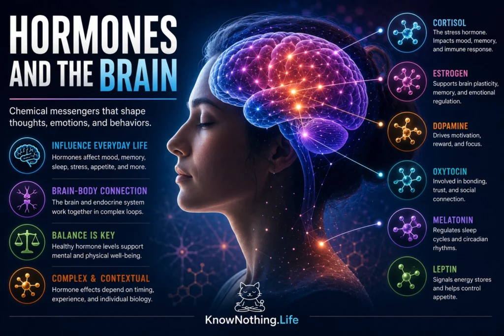
Hormones are chemical messengers that coordinate activity across the body, but their influence extends far beyond metabolism, growth, and reproduction. They also affect attention, memory, motivation, sleep, appetite, emotion, and social behavior. The brain is both a target of hormonal signals and one of their principal regulators. Through the hypothalamus and pituitary gland, neural circuits translate information about stress, light, nutrition, temperature, and reproduction into endocrine responses that can reach nearly every organ.
Hormonal effects depend on whether brain cells possess the appropriate receptors. Steroid hormones such as cortisol, estradiol, progesterone, and testosterone can enter cells and bind receptors that influence gene expression. Other hormones act through receptors on cell membranes, producing faster changes in electrical activity, neurotransmitter release, or intracellular signaling. Bruce McEwen, Joseph Weiss, and Lloyd Schwartz demonstrated in their 1968 study Selective Retention of Corticosterone by Limbic Structures in Rat Brain that corticosterone was strongly retained in the hippocampus. The finding helped establish that hormones produced outside the brain could act directly on regions associated with learning, emotion, and memory.
Stress Hormones and the Adaptive Response
The hypothalamic-pituitary-adrenal axis coordinates a major part of the body’s response to stress. When the brain detects a threat or significant demand, the hypothalamus releases corticotropin-releasing hormone. This stimulates the pituitary gland to secrete adrenocorticotropic hormone, which travels through the bloodstream and prompts the adrenal cortex to release cortisol. Cortisol helps mobilize energy, modifies immune activity, and shifts attention toward information that may be important for immediate survival.
Cortisol’s effects on the brain are complex. A temporary increase can support alertness and strengthen the consolidation of emotionally significant experiences, while stress occurring during memory retrieval can make stored information more difficult to access. Persistently elevated cortisol may interfere with hippocampal and prefrontal functions. Sonia Lupien and colleagues reported in 1998 that older adults whose cortisol levels increased over several years showed reduced hippocampal volume and poorer hippocampus-dependent memory. The study did not prove that cortisol alone caused every difference, but it demonstrated a close relationship between long-term hormonal patterns, brain structure, and cognitive performance.
Sex Hormones and Neural Plasticity
Estradiol, progesterone, and testosterone act on the brain throughout life rather than only controlling reproductive organs. Their receptors are distributed across the hypothalamus, amygdala, hippocampus, cerebral cortex, and other regions. These hormones contribute to sexual development and reproductive behavior, but they can also influence mood, reward, pain sensitivity, motivation, and synaptic plasticity. Testosterone may act directly through androgen receptors or be converted into estradiol by the enzyme aromatase, meaning that the effect of a circulating hormone depends partly on how it is processed within a particular tissue.
Research by Catherine Woolley and Bruce McEwen showed that estradiol can produce measurable changes in hippocampal circuitry. Their 1992 study Estradiol Mediates Fluctuation in Hippocampal Synapse Density During the Estrous Cycle in the Adult Rat found that higher estradiol levels were associated with increased synapse density in hippocampal CA1 neurons. Later experiments showed that estradiol increased dendritic spine density through a mechanism requiring NMDA receptor activation and enhanced the sensitivity of hippocampal neurons to NMDA-mediated input. These findings demonstrated that a hormone associated with reproduction can also remodel neural connections involved in learning and memory.
Thyroid Hormones and Brain Development
Thyroid hormones are essential for normal development of the nervous system. During prenatal and early postnatal life, they influence neuronal differentiation, migration, axonal growth, synapse formation, and myelination. Severe untreated congenital hypothyroidism can therefore cause profound intellectual and neurological impairment. Newborn screening programs transformed outcomes by allowing thyroid hormone replacement to begin before prolonged deficiency could disrupt sensitive stages of brain development.
Studies following children with congenital hypothyroidism have shown that early treatment is associated with substantially better cognitive development than the outcomes historically observed without treatment. Research comparing treatment schedules has found that the timing of levothyroxine replacement can be especially important for later intellectual performance. Maternal thyroid function also matters during pregnancy because the developing fetal brain depends partly on maternal hormone supply. A prospective cohort study found that both unusually low and unusually high maternal thyroid hormone levels were associated with differences in childhood IQ and brain morphology, illustrating that hormonal development depends on appropriate timing and range rather than simply maximizing the amount of a hormone.
Metabolic Hormones and Appetite
The brain continuously receives hormonal information about the body’s energy condition. Insulin communicates information related to blood glucose and nutrient storage, while hormones released by the digestive system and adipose tissue influence hunger, satiety, energy expenditure, and food reward. These signals converge in hypothalamic and brainstem circuits but also interact with memory, stress, sensory cues, and learned expectations. Eating is therefore controlled by a distributed regulatory system rather than a single hunger center.
The discovery of leptin revealed that fat tissue functions as an endocrine organ. In 1995, Louis Tartaglia and colleagues identified and cloned the leptin receptor in Identification and Expression Cloning of a Leptin Receptor, OB-R. The receptor was found in the hypothalamus and several other tissues. Subsequent experiments showed that leptin activates neurons in hypothalamic regions involved in energy regulation and stimulates the intracellular signaling protein STAT3. Leptin generally informs the brain about stored energy, but many people with obesity have high leptin levels and reduced responsiveness to the signal. This illustrates a recurring neuroendocrine principle: hormone concentration matters, but receptor sensitivity and downstream signaling are equally important.
Oxytocin, Vasopressin, and Social Behavior
Oxytocin and vasopressin are produced by hypothalamic neurons and released both into the bloodstream and within the nervous system. In the body, oxytocin supports uterine contractions and milk ejection, while vasopressin regulates water balance and blood pressure. Within the brain, both peptides can influence social recognition, parental behavior, defensive responses, affiliation, and attachment. Their effects depend on receptor location, developmental history, biological sex, and social context, making descriptions of oxytocin as a universal “love hormone” misleading.
Prairie vole studies became influential because these animals can form enduring social bonds. Jessie Williams, Thomas Insel, C. Sue Carter, and colleagues reported in 1994 that central oxytocin administration facilitated partner preference formation in female prairie voles, while blocking oxytocin receptors prevented the effect. Other experiments found that vasopressin signaling contributed to partner preference and selective aggression in males. Introducing additional vasopressin receptors into reward-related forebrain regions also increased affiliative behavior. More recent genetic research has complicated the original model by showing that prairie voles without functioning oxytocin receptors can still form pair bonds, indicating that attachment emerges from overlapping and partially compensatory neural systems.
Melatonin and the Biological Clock
Melatonin is produced mainly by the pineal gland according to signals from the brain’s circadian clock. Its secretion normally rises during biological night, providing the nervous system and other organs with information about environmental time. Melatonin receptors are found in the suprachiasmatic nucleus, the hypothalamic region that coordinates daily rhythms. Through these receptors, melatonin can influence sleep timing and help adjust the phase of circadian activity.
Experiments involving genetically altered mice have clarified the importance of receptor-mediated melatonin signaling. In animals lacking the MT1 receptor, melatonin failed to produce the normal shift in circadian activity seen in control mice. Other research has shown that nightly melatonin exposure changes the responsiveness of MT2 receptors in the suprachiasmatic nucleus. These findings demonstrate that melatonin does not simply produce sleepiness. It participates in a timing system that coordinates sleep, temperature, metabolism, attention, and endocrine secretion with the expected cycle of day and night.
Hormones Influence Behavior Without Determining It
Hormones do not issue fixed commands or produce identical behavior in everyone. The same cortisol increase may improve attention in one situation and disrupt memory in another. Testosterone is not a simple aggression chemical, and oxytocin does not make every social encounter trusting or affectionate. Effects depend on hormone dose, receptor distribution, previous experience, interacting neurotransmitters, developmental stage, and the meaning of the situation to the individual. Experimental testosterone administration, for example, has produced stronger aggressive responses primarily among men who were already dominant or impulsive, rather than creating a universal effect.
The most accurate view is that hormones participate in feedback loops connecting the brain, body, behavior, and environment. Experience changes endocrine activity, hormones modify neural processing, and behavior creates new experiences that feed back into the system. Hormones help the brain assign biological priorities—when to seek food, conserve energy, respond to danger, sleep, reproduce, or approach other people. They are not hidden forces controlling the mind from outside. They are part of the biological machinery through which the brain interprets the body’s condition and adapts to a changing world.


