
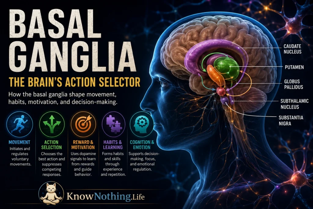
The basal ganglia are a group of interconnected structures located deep within the cerebral hemispheres and upper brainstem. Their major components include the caudate nucleus, putamen, globus pallidus, subthalamic nucleus, and substantia nigra. The caudate and putamen together form the dorsal striatum, which receives extensive information from the cerebral cortex. Related ventral structures, including the nucleus accumbens and ventral pallidum, connect basal-ganglia circuitry with motivation, emotion, and reward. Rather than directly commanding muscles, these nuclei influence which actions are selected, how forcefully they are performed, and when competing behaviors should be suppressed.
Basal-ganglia function extends beyond physical movement. Parallel circuits link these structures with motor, oculomotor, associative, and limbic regions of the cortex. Anatomical tracing by James Hoover and Peter Strick demonstrated that the internal globus pallidus contains separate output channels projecting through the thalamus toward distinct cortical motor areas. This organization helps explain how similar circuit principles can influence reaching, eye movements, decision-making, motivation, and learned behavioral routines without combining every function into one undifferentiated pathway.
The Main Structures and Circuit Loops
The striatum is the principal entry point for information arriving from the cerebral cortex. Most of its projection neurons are inhibitory spiny projection neurons, which remain relatively quiet until suitable combinations of cortical and thalamic input activate them. The internal globus pallidus and substantia nigra pars reticulata serve as major output structures. Their neurons normally fire continuously and inhibit targets in the thalamus and brainstem. Movement-related signals can alter this inhibition, allowing selected motor or cognitive programs to influence downstream systems.
These circuits operate as loops. Cortical activity enters the striatum and other basal-ganglia structures, passes through pallidal or nigral output nuclei, reaches the thalamus, and returns to related cortical regions. The loops are partly segregated but not completely isolated. Motor information may interact with reward, attention, and context at several levels. The substantia nigra pars compacta supplies dopamine to the striatum, modifying the responsiveness and plasticity of its circuits. The basal ganglia therefore do not create movement independently; they regulate activity already being prepared across wider cortical, thalamic, and brainstem networks.
Direct, Indirect, and Hyperdirect Pathways
The traditional circuit model distinguishes direct and indirect pathways. Direct-pathway striatal neurons project to the internal globus pallidus and substantia nigra pars reticulata. Because these connections are inhibitory, their activation can reduce the output nuclei’s continuing inhibition of thalamic or brainstem targets. This disinhibition can help a selected action influence motor systems. Indirect-pathway neurons project first to the external globus pallidus, which communicates with the subthalamic nucleus and output nuclei. Through this longer route, indirect-pathway activity can increase inhibitory basal-ganglia output and constrain behavior.
Optogenetic research provided strong causal evidence that the pathways can produce opposing motor effects. Anatol Kreitzer’s group selectively activated direct- or indirect-pathway neurons in parkinsonian mice. Direct-pathway stimulation improved movement, while indirect-pathway stimulation increased freezing and reduced locomotion. The result supported the broad facilitatory and suppressive roles assigned to the pathways, although it did not establish that they always operate separately or control every action through a simple “go” and “stop” division.
The hyperdirect pathway carries cortical signals rapidly to the subthalamic nucleus, which can increase activity in basal-ganglia output nuclei. This route may help interrupt an automatic response when circumstances suddenly require greater control. Masaki Isoda and Okihide Hikosaka recorded from monkey subthalamic neurons during a task requiring a switch from an habitual eye movement to a deliberately controlled alternative. Subthalamic activity was associated with suppressing the automatic response and enabling the new action.
Action Selection and Movement Regulation
One influential view describes the basal ganglia as an action-selection system. At any moment, the brain may contain several potential movements or behavioral strategies. Basal-ganglia circuits can help promote the program most appropriate to the current goal while reducing interference from alternatives. This function applies not only to choosing whether to reach left or right, but also to selecting thought sequences, emotional responses, eye movements, and learned routines.
Modern findings have complicated the idea that direct- and indirect-pathway neurons merely oppose each other. Guohong Cui and colleagues monitored both populations while mice initiated learned actions. Activity increased in direct and indirect pathways at approximately the same time, and their combined patterns predicted specific upcoming movements. One interpretation is that direct-pathway activity supports the chosen action while indirect-pathway activity constrains competing or inappropriate components. The two pathways may therefore cooperate to sharpen behavior rather than alternately switching the entire motor system on and off.
The basal ganglia also influence movement vigor—the speed, force, and energy invested in an action. Dopamine depletion can make movements slow and difficult to initiate without eliminating the person’s understanding of what movement is required. Conversely, excessive or poorly regulated basal-ganglia activity can contribute to unwanted movements. These observations suggest that the system helps determine not only which action is selected but how strongly its expected benefit justifies the effort required to perform it.
Dopamine, Reward, and Reinforcement Learning
Dopamine is essential to basal-ganglia learning and movement, but it should not be described simply as a pleasure chemical. Dopamine released in the striatum modifies synaptic plasticity, neuronal excitability, motivation, and the balance among competing actions. Direct-pathway neurons generally express more D1-family dopamine receptors, while many indirect-pathway neurons express D2-family receptors. Dopamine can therefore influence the two populations differently, although its effects depend on firing patterns, local circuitry, and behavioral state.
Wolfram Schultz and colleagues recorded from dopamine neurons while monkeys learned relationships between cues and rewards. Unexpected rewards produced a brief increase in firing. Once a cue reliably predicted the reward, the response shifted from the reward itself to the predictive cue. If an expected reward failed to appear, dopamine activity decreased around the time it should have occurred. Schultz interpreted these patterns as a reward-prediction error: a teaching signal representing the difference between what was expected and what actually happened.
Prediction-error signals can strengthen actions whose outcomes are better than expected and weaken those followed by disappointing results. Through repeated experience, corticostriatal connections become more likely to support choices that have produced useful outcomes in similar situations. Dopamine also affects willingness to expend effort, linking reinforcement learning with action vigor. Basal-ganglia learning is consequently not separate from motor control; it continually reshapes which actions are selected and how readily they are performed.
Habit Formation and Automatic Behavior
Early in learning, a task may require conscious attention to every step. With practice, separate actions can become grouped into larger behavioral units that are executed more efficiently. The striatum is central to this transition from deliberate action to habitual performance. Habits reduce cognitive demands, but they can also become resistant to changes in goals or consequences. Basal-ganglia circuits therefore help make skilled behavior efficient while creating the possibility that routines may persist after they are no longer useful.
Mark Jog, Yasuo Kubota, and Ann Graybiel recorded from striatal neurons as rats learned a T-maze task. Neural activity changed widely during acquisition, eventually becoming concentrated near the beginning and end of the practiced sequence. The pattern remained stable during later performance, suggesting that the striatum had reorganized the individual actions into a unified behavioral “chunk.”
Habit learning does not mean that the striatum becomes inactive once a routine is established. Recordings from trained animals show that parts of the dorsolateral striatum continue to encode position, speed, timing, and task context during habitual sequences. The basal ganglia may therefore contribute both to creating automatic routines and to constraining their accurate execution. When circumstances change, interactions with prefrontal and associative circuits can help replace a habitual response with a more flexible, goal-directed choice.
Parkinson’s Disease, Huntington’s Disease, and Dystonia
Parkinson’s disease is strongly associated with the degeneration of dopamine-producing neurons in the substantia nigra pars compacta. Reduced striatal dopamine disrupts basal-ganglia activity and contributes to bradykinesia, rigidity, resting tremor, and difficulty initiating movement. The disorder cannot be explained by firing rates alone; abnormal synchrony, bursting, and oscillatory patterns also develop across the network. Recordings from parkinsonian monkeys have found increased and rhythmically bursting activity in the subthalamic nucleus, demonstrating that dopamine loss alters the timing as well as the overall level of basal-ganglia output.
Huntington’s disease produces a different progression. Degeneration affects striatal projection neurons, with early vulnerability in populations contributing to the indirect pathway. Reduced suppression of competing actions is associated with chorea, the involuntary movements characteristic of early disease. Quantitative studies of human tissue have demonstrated differential loss among striatal projection systems, while later degeneration becomes more extensive and may contribute to rigidity and slowed movement.
Dystonia involves sustained or intermittent muscle contractions that produce abnormal postures or repetitive movements. It is increasingly understood as a network disorder involving basal-ganglia, cerebellar, cortical, and sensory systems rather than a defect in one nucleus. A randomized controlled trial found that stimulation of the internal globus pallidus improved symptoms in people with primary generalized or segmental dystonia, showing that altering basal-ganglia output can reorganize movement across the wider motor network.
Deep Brain Stimulation and the Modern View
Deep brain stimulation delivers electrical pulses through implanted electrodes, commonly targeting the subthalamic nucleus or internal globus pallidus in advanced Parkinson’s disease. A large prospective crossover study found that stimulation of either target improved motor function and activities of daily living when medication alone no longer provided adequate control. DBS does not simply destroy or silence a nucleus. Its effects appear to involve changes in pathological firing patterns, axonal signaling, network synchrony, and information flow across basal-ganglia–thalamocortical circuits.
The modern view of the basal ganglia is therefore more complex than the original diagram of opposing pathways. Direct, indirect, and hyperdirect circuits can operate together; dopamine contributes to movement, learning, effort, and prediction; and different loops influence cognitive and emotional behavior as well as muscles. The basal ganglia do not issue detailed commands for every joint. They help the nervous system choose among possibilities, reinforce successful strategies, organize practiced sequences, and regulate the intensity with which an action is expressed. Their function is best understood as adaptive control over behavior—shaping what the brain does next and how readily it does it.


