
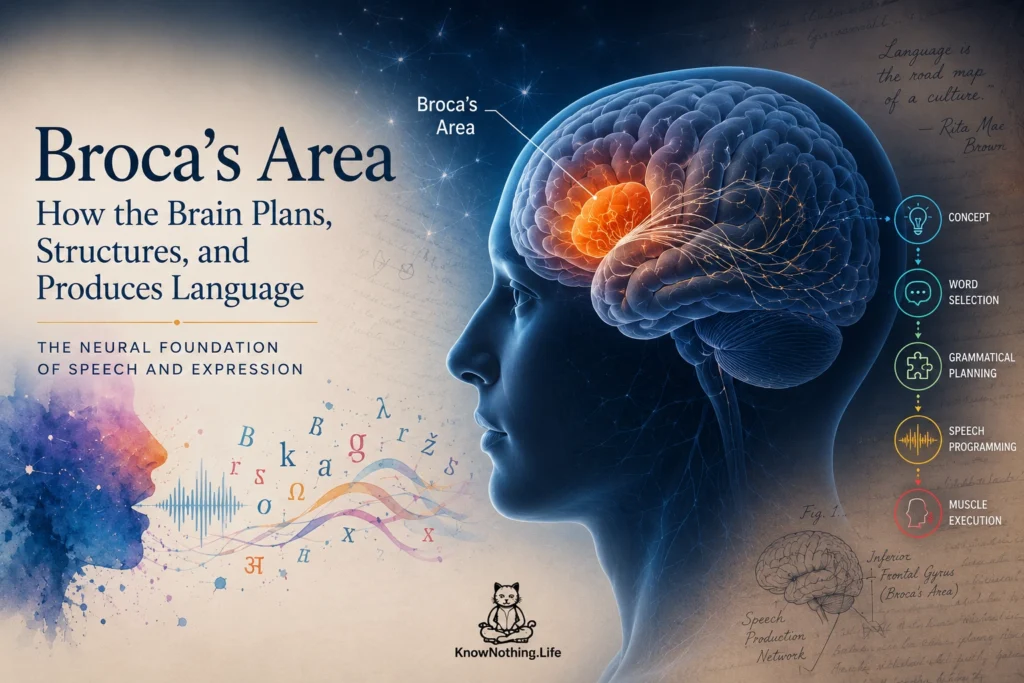
Broca’s area is a region of the left frontal lobe traditionally associated with speech production and language. It lies within the inferior frontal gyrus, most commonly encompassing the pars opercularis and pars triangularis, which roughly correspond to Brodmann areas 44 and 45. Textbooks long described it as the brain’s speech center: damage caused slow, broken speech while comprehension remained relatively intact. That account is too simple. The region participates in several stages of language processing and sits beside systems involved in attention, cognitive control, and action planning.
Contemporary research treats Broca’s area less as a single-purpose module and more as neighboring cortical territories with different functional profiles. Some parts respond selectively during language, while adjacent areas become active during demanding nonlinguistic tasks. Their boundaries also vary across individuals, making anatomy alone an imperfect guide to function. Broca’s area is therefore best understood as a frontal component of a distributed language network rather than the single place where speech or grammar is stored.
Paul Broca and the Discovery of Language Localization
The region takes its name from French physician Pierre Paul Broca. In 1861, Broca presented the case of Louis Victor Leborgne, a man who had lost nearly all propositional speech and was known by the syllable he repeatedly produced: “tan.” After Leborgne died, Broca found extensive damage in the left frontal lobe. He soon described another patient with a similar disorder and left-sided frontal damage. These observations strengthened the then-controversial claim that complex mental functions could be localized to particular parts of the cerebral cortex.
Modern imaging of the preserved brains of Broca’s patients complicated that story. Nina Dronkers and colleagues used high-resolution magnetic resonance imaging to reconstruct their lesions and found that the damage extended beyond the cortical region now labeled Broca’s area. Leborgne’s lesion involved frontal, parietal, temporal, insular, and deep white-matter structures, while the second patient also had more extensive injury than surface inspection revealed. Broca’s observations were transformative, but his patients did not prove that injury to one small patch produces the full syndrome later called Broca’s aphasia.
Anatomy and Functional Organization
Broca’s area occupies the posterior inferior frontal gyrus, above the lateral fissure and in front of regions controlling movements of the mouth and face. The pars opercularis, associated approximately with area 44, lies posteriorly; the pars triangularis, associated with area 45, lies anteriorly. Cellular architecture, connectivity, and imaging indicate that these territories are not interchangeable. Area 44 is often linked to phonological processing, sequencing, and speech-related planning, while area 45 frequently contributes to controlled retrieval and selection among competing linguistic representations.
Even this subdivision is not sufficiently precise. Evelina Fedorenko, John Duncan, and Nancy Kanwisher used functional localization within individual participants and found language-selective and domain-general regions positioned side by side inside the broad territory called Broca’s area. Group-averaged maps can blur these systems because their borders differ across people. This helps explain why the region has been associated with language, working memory, action, music, and cognitive control: different tasks may activate distinct but adjacent populations of cortex.
From Words to Articulation
Speaking requires the brain to move from intended meaning to selected words, grammatical forms, sound patterns, and coordinated movements of the lips, tongue, larynx, and respiratory system. Broca’s area is active during this process, but it does not directly command every speech muscle. Evidence instead suggests that it helps prepare and sequence linguistic and articulatory information before motor cortex executes the movements. A person with aphasia may therefore struggle to formulate speech even though the required muscles are not paralyzed.
Adeen Flinker and colleagues recorded electrical activity directly from the cortex of people preparing to speak. Activity moved from temporal regions to Broca’s area and then to motor cortex during word production. Broca’s area was strongly engaged before articulation but became less active once speech began, while motor cortex remained active during execution. The findings supported a coordinating or preparatory role rather than the idea that Broca’s area continuously drives articulation. Stimulation research likewise suggests that disrupting it can prevent speech initiation without eliciting the muscle responses produced by stimulating primary motor cortex.
Grammar, Phonology, and Linguistic Sequencing
Broca’s area has long been linked to grammar because people with nonfluent aphasia may omit function words and grammatical endings, producing short expressions composed mainly of nouns and verbs. Comprehending simple sentences may remain relatively strong, while unusual word order or long-distance relationships can create difficulty. These observations encouraged theories that the left inferior frontal gyrus performs syntactic operations. Modern evidence indicates that it contributes to sentence processing, but grammar depends on cooperation between frontal and temporal language regions rather than one isolated center.
Ned Sahin and colleagues used intracranial electrodes to measure activity within Broca’s area while participants produced inflected words. Within several hundred milliseconds, the same small region showed patterns associated first with identifying a word, then with applying grammatical information, and later with preparing its sound structure. The study demonstrated rapid sequential processing of lexical, grammatical, and phonological information within frontal language cortex. It did not show that these functions exist only there. The region appears to organize and transform representations received from a wider network, especially when linguistic material must be ordered or converted into a planned response.
Broca’s Area Within the Language Network
Language depends on extensive connections between frontal and temporal cortex. Ventral pathways help connect spoken or written forms to meaning, while dorsal pathways support sound-to-motor mapping, repetition, sequencing, and aspects of complex sentence processing. Broca’s area communicates with posterior temporal, inferior parietal, premotor, supplementary motor, insular, basal ganglia, and cerebellar systems. Damage to white matter connecting these regions can impair language even when important cortical areas remain preserved.
This network perspective explains why a small lesion confined to Broca’s area does not always cause lasting Broca’s aphasia. A study of chronic stroke found that the classic syndrome was most strongly associated with damage involving both frontal and posterior language regions. Another study of 134 stroke survivors reported that damage limited to Broca’s area did not independently predict poor long-term speech production once injury to surrounding systems was considered. In acute stroke, frontal ischemia may produce severe nonfluency, but some patients improve as surviving parts of the network reorganize.
Broca’s Aphasia and Clinical Symptoms
Broca’s aphasia, also called nonfluent or expressive aphasia, commonly involves slow, effortful, reduced speech. People may know what they wish to communicate but struggle to retrieve words, form grammatical sentences, or produce fluent sequences. Speech may consist mainly of content words, with articles, prepositions, auxiliary verbs, and inflections omitted. Naming, repetition, reading, and writing may also be affected. Comprehension is often better preserved than production, but it may be impaired for grammatically complex sentences.
The syndrome must be distinguished from apraxia of speech and dysarthria. Apraxia is a disorder of planning speech movements, while dysarthria results from impaired muscular control. These disorders can accompany aphasia because strokes rarely respect textbook boundaries, but they are not identical to language impairment. Assessment therefore examines spontaneous speech, naming, repetition, comprehension, reading, writing, articulation, and nonverbal cognition. The individual pattern matters more than the broad label and helps determine which processes therapy should target.
Recovery, Plasticity, and the Modern View
Recovery after left frontal injury depends on lesion size, damaged pathways, age, health, and the integrity of the remaining network. Speech-language therapy may focus on word retrieval, sentence construction, articulatory planning, reading, writing, and conversation. Improvement is usually supported by surviving tissue around the lesion and other left-hemisphere language regions, although right-hemisphere systems may assist under some conditions. Recovery does not mean that language simply moves into a mirror image of Broca’s area; it reflects changing cooperation across networks.
The modern account preserves Broca’s central insight while rejecting the strongest version of localization. The left frontal lobe is critically involved in language, and the region bearing his name contributes to selecting, structuring, sequencing, and preparing linguistic information for expression. Yet it is neither a solitary speech organ nor a uniform patch of cortex. Broca’s area contains neighboring specialized and general-purpose systems, works through long-range connections, and changes its contribution according to the task. Its history illustrates a broader lesson: complex abilities can depend on identifiable regions without being confined to them.


