
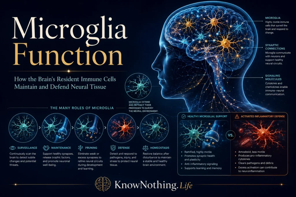
Microglia are specialized immune cells distributed throughout the brain and spinal cord. They are often called the resident macrophages of the central nervous system because they detect danger, engulf unwanted material, coordinate inflammatory responses, and assist tissue repair. That description is incomplete, however. Microglia also perform essential functions in the healthy brain, including monitoring neural activity, shaping developing circuits, supporting plasticity, and clearing dying cells before they release damaging contents.
Unlike many immune cells that are continually replaced from bone marrow, most microglia arise early in embryonic development. In a landmark 2010 study, Florent Ginhoux and colleagues used lineage tracing in mice to show that adult microglia descend from primitive myeloid progenitors appearing before embryonic day eight. These cells enter the developing nervous system, establish long-lived populations, and largely maintain themselves through local renewal. Their unusual origin helps explain why microglia differ molecularly from circulating monocytes and other tissue macrophages.
Continuous Surveillance of the Brain
The older phrase “resting microglia” suggested that these cells remain inactive until infection or injury occurs. Live imaging overturned that view. In 2005, Axel Nimmerjahn, Frank Kirchhoff, and Fritjof Helmchen observed microglia in the living mouse brain and found that their cell bodies remained relatively stable while their thin processes constantly extended, retracted, and sampled nearby tissue. Microglia are therefore better described as surveying or homeostatic cells. They continually inspect extracellular conditions, blood vessels, synapses, and neighboring glia for signs of change.
This surveillance shifts with brain state. Research led by Yong Liu in 2019 showed that microglial processes covered less territory in awake mice than during anesthesia or reduced neuronal activity. Norepinephrine released during wakefulness suppressed process extension, linking microglial behavior to arousal. Their movements are therefore regulated by neuronal and neuromodulatory signals rather than occurring at one fixed rate.
Responding to Injury and Infection
Damaged cells rapidly release ATP and related nucleotides into surrounding tissue. In 2005, Dimitrios Davalos and colleagues showed that ATP acts as an early distress signal directing microglial processes toward a small brain injury. Sharon Haynes and colleagues later demonstrated that the P2Y12 receptor is critical for this movement: microglia lacking it retained ordinary baseline motility but failed to orient efficiently toward extracellular nucleotides.
Once engaged, microglia can engulf dead cells, microbes, damaged myelin, abnormal protein deposits, and cellular fragments. They also release cytokines, chemokines, reactive molecules, and growth factors that influence neurons, astrocytes, blood vessels, and infiltrating immune cells. These actions can contain infection and remove hazardous material, but they require tight control. Too little response may allow damage to spread, whereas prolonged inflammation can injure healthy neurons and synapses.
Clearing Cells and Maintaining Tissue
Phagocytosis—the ingestion and digestion of unwanted material—is a central microglial function and is not limited to disease. During adult hippocampal neurogenesis, many newly generated cells die before becoming integrated neurons. Amanda Sierra and colleagues reported in 2010 that microglia rapidly engulf these apoptotic cells while producing little detectable inflammation. Efficient clearance prevents cellular contents from spilling into nearby tissue and allows neurogenic regions to remain stable despite continuous cell birth and death.
Microglial clearance also contributes to recovery after injury. Cells must distinguish debris that should be removed from stressed tissue that may still recover. Damaged cells can display molecular signals encouraging engulfment, while healthy cells express cues that discourage it. If these recognition systems become unbalanced, microglia may remove viable neural structures or fail to clear harmful material. Phagocytosis is therefore a selective decision process rather than simple waste disposal.
Shaping Synapses During Development
The developing brain initially produces more synaptic connections than it retains. Neural activity strengthens useful pathways, while weaker or poorly coordinated connections are eliminated. Beth Stevens and colleagues showed in 2007 that proteins from the classical complement system, especially C1q and C3, participate in this refinement. Complement molecules localize to selected synapses, effectively marking them for removal through an immune mechanism adapted for brain development.
Rosa Paolicelli and colleagues demonstrated in 2011 that microglia engulf synaptic material and are required for normal postnatal pruning in mice. A later study led by Dorothy Schafer found that microglial removal of retinal inputs depended on neural activity and complement receptor signaling. These findings changed the understanding of microglia from emergency responders into active architects of neural circuits. Pruning must remain precise: too little can preserve inefficient connections, while excessive elimination may weaken useful networks.
Supporting Plasticity and Learning
Microglia do not only remove synapses; they may also help create and stabilize them. In 2013, Cody Parkhurst and colleagues temporarily depleted microglia in mice and observed impaired performance on several learning tasks together with reduced formation of new synapses during motor learning. Removing brain-derived neurotrophic factor from microglia produced similar effects, suggesting that microglial signaling can support experience-dependent circuit remodeling.
Microglia also make brief contacts with functioning synapses. Hiroaki Wake and colleagues reported that these contacts were influenced by neuronal activity and became more prolonged after brain injury or reduced blood flow. Such interactions may allow microglia to evaluate synaptic condition and respond to local changes. They do not perform neuronal computation, but they help maintain the environment in which neural networks communicate and adapt.
Microglial States and Functional Diversity
Microglia change their shape, metabolism, receptors, and gene expression according to local conditions. The distinction between “inactive” and “activated” microglia is therefore too crude. The once-popular M1/M2 model, which classified cells as either inflammatory or repair-oriented, also fails to capture the diversity observed in living tissue. One microglial population may simultaneously perform protective and potentially damaging functions, and its behavior may change as a disease progresses.
Single-cell sequencing has revealed populations associated with development, aging, injury, tumors, demyelination, and neurodegeneration. In 2017, Hadas Keren-Shaul and colleagues identified disease-associated microglia in mouse models of Alzheimer’s disease and other disorders. Their transition involved a program partly dependent on TREM2, a receptor associated with lipid sensing and phagocytosis. These states are not automatically beneficial or harmful; their effects depend on timing and context.
Microglia in Neurodegenerative Disease
In Alzheimer’s disease, microglia gather around amyloid plaques, engulf material, and alter inflammatory and metabolic pathways. Human genetic evidence involving TREM2 and other microglial genes indicates that these cells influence disease risk. Yet their role is not simple: microglia may compact plaques and limit their spread while also releasing inflammatory mediators or removing stressed synapses. Soyon Hong and colleagues showed in 2016 that complement proteins and microglia mediated early synaptic loss in mouse models of Alzheimer’s disease.
Similar tensions appear in Parkinson’s disease, multiple sclerosis, amyotrophic lateral sclerosis, Huntington’s disease, stroke, and traumatic injury. Microglia can clear toxic proteins and damaged myelin, support remyelination, and promote repair, but persistent activation may sustain oxidative stress or disrupt neuronal communication. The outcome depends on disease stage, brain region, genetics, age, and signals from neighboring cells.
Therapeutic Possibilities and Remaining Questions
Microglia are attractive therapeutic targets because they influence many stages of neurological disease. Experimental strategies include altering TREM2 signaling, regulating complement, blocking selected cytokines, modifying metabolism, or temporarily reducing microglial populations through inhibition of colony-stimulating factor 1 receptor signaling. In 2014, Michelle Elmore and colleagues showed that CSF1R inhibition could eliminate most microglia in the adult mouse brain and that the population rapidly returned after treatment stopped. This revealed unexpected regenerative capacity, but it did not establish that depletion would be safe or useful in people.
The central challenge is preserving normal microglial functions while limiting harmful ones. Broad suppression could weaken immune defense, debris removal, circuit maintenance, and tissue repair. Future treatments will probably need to target a particular state, pathway, brain region, or disease stage rather than treating every microglial cell as identical. Human tissue studies, advanced imaging, organoids, and single-cell methods are helping researchers determine which findings from animal models translate to human disease.
Microglia are not merely immune cells waiting for the brain to become diseased. They are lifelong participants in development, surveillance, synaptic refinement, learning, defense, and repair. Their flexibility is their greatest strength and also the reason they can become involved in pathology. Understanding microglial function requires moving beyond the question of whether these cells are “good” or “bad” and asking which state they occupy, what signals created it, and what that state is doing within a particular neural environment.


