
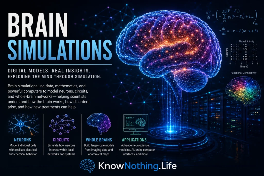
Brain simulations are computer-based models designed to reproduce selected structures, signals, or functions of the nervous system. Some represent the electrical activity of individual neurons in great biological detail, while others describe the average behavior of entire brain regions. Researchers use these simulations to investigate how cellular properties, synaptic connections, neural dynamics, and anatomical networks might produce perception, memory, movement, decision-making, or disease. A simulation can also allow scientists to manipulate variables that would be difficult, expensive, or unethical to change directly in a living brain.
The phrase “brain simulation” can be misleading because no existing model reproduces every neuron, synapse, molecule, developmental process, and bodily interaction of a complete human brain. Every simulation is selective. It includes the mechanisms considered important for a particular question and simplifies or excludes others. The scientific value of a model therefore does not depend only on its size. A smaller, interpretable simulation that makes accurate predictions may be more useful than a massive model whose assumptions are poorly constrained.
Choosing the Right Level of Detail
Brain activity unfolds across many levels of organization. Ion channels shape the voltage of a single membrane, dendrites transform synaptic inputs, local circuits coordinate excitation and inhibition, and long-range pathways connect specialized regions. Simulating all these processes simultaneously creates enormous computational and experimental challenges. Researchers must decide whether neurons should be represented as detailed multicompartment cells, simplified spiking units, firing-rate populations, or abstract dynamical systems. Each level answers different questions.
Eugene Izhikevich and Gerald Edelman illustrated the scale of this challenge in their 2008 model of mammalian thalamocortical systems. The simulation combined human diffusion-imaging data, cortical organization derived from animal studies, 22 modeled neuronal types, approximately one million multicompartment spiking neurons, and nearly half a billion synapses. It demonstrated that large-scale anatomical and physiological features could be integrated within one system, but it also revealed a continuing limitation of ambitious brain simulations: missing human data often require assumptions or information drawn from different species and experimental conditions.
Reconstructing Cortical Microcircuits
One major approach attempts to reconstruct small pieces of brain tissue in high biological detail. Henry Markram and colleagues’ 2015 study, “Reconstruction and Simulation of Neocortical Microcircuitry,” created a first-draft digital model of a small volume of juvenile rat somatosensory cortex. The reconstruction contained roughly 31,000 neurons and approximately 37 million synapses, with cellular forms and electrical properties constrained by anatomical and patch-clamp data. The model reproduced several patterns of spontaneous and evoked network activity and allowed researchers to explore how cellular diversity and connectivity shape cortical dynamics.
Other projects prioritize scale and reproducibility over cellular detail. Tobias Potjans and Markus Diesmann developed a full-scale spiking model of a cortical microcircuit containing about 77,000 simplified neurons organized across four cortical layers. Its integrated connectivity map produced asynchronous, irregular activity and layer-specific firing rates resembling experimental observations. Such models show that realistic collective behavior can emerge even when individual neurons are represented simply, provided that population sizes, connectivity patterns, synaptic strengths, and delays are appropriately constrained.
Integrating Large Experimental Datasets
Modern brain simulations increasingly rely on standardized, open datasets rather than isolated measurements gathered from different laboratories. Yazan Billeh and colleagues integrated anatomical, physiological, and functional data into a multiscale model of awake mouse primary visual cortex. The model included more than 230,000 neurons and was implemented using both biophysically detailed cells and simplified point neurons. The two versions shared the same network connectivity and produced similar firing-rate distributions for the questions studied, showing that additional biological detail does not automatically change every network-level prediction.
This finding illustrates a central principle of simulation design: complexity should be justified by the phenomenon being investigated. Detailed dendritic and ion-channel models may be essential when studying how drugs alter cellular excitability, but simplified neurons may be sufficient for examining population firing, signal propagation, or network stability. Open access to model code, parameters, and experimental data is also critical because simulations can be difficult to reproduce when their assumptions are scattered across publications or embedded within undocumented software.
Simulating Cognition and Behavior
Many large simulations reproduce neural activity without demonstrating how that activity generates useful behavior. The Semantic Pointer Architecture Unified Network, known as Spaun, was designed to bridge this divide. Chris Eliasmith and colleagues constructed a model containing approximately 2.5 million spiking neurons organized into systems associated with vision, working memory, action selection, and motor output. Spaun received visual information and generated responses through a physically modeled arm while completing eight tasks, including image recognition, memory, counting, and simple reasoning.
Spaun was not intended to reproduce every feature of the human brain. Its importance came from connecting neural representations and anatomical constraints with several forms of behavior inside one unified architecture. The model also displayed some human-like patterns of errors and performance. Functional simulations of this kind allow scientists to ask how the same neural mechanisms might support several tasks rather than building an unrelated model for each behavior. They also reveal how damage to simulated components could influence performance across multiple cognitive functions.
Whole-Brain Network Models
At the opposite end of the spectrum from cellular reconstruction are whole-brain network simulations. These models divide the brain into regions and connect them using structural information obtained through techniques such as diffusion MRI. Each region is represented by a mathematical model of local population activity. The simulation then examines how neural dynamics travel across the structural network and generate patterns resembling electroencephalography, magnetoencephalography, or functional MRI signals.
Christopher Honey and colleagues showed that structural connectivity strongly shapes simulated resting-state functional connectivity, although indirect connections and neural dynamics were required to explain relationships between regions that lacked direct anatomical links. The Virtual Brain platform, introduced by Paula Sanz Leon, Petra Ritter, and their colleagues, expanded this approach by allowing researchers to simulate large-scale brain-network activity using biologically informed connectivity and regional dynamics. These models provide a framework for testing how anatomical structure constrains the changing patterns measured through neuroimaging.
Personalized Simulations and Virtual Brain Twins
Whole-brain models can be personalized by constructing their structural networks from an individual patient’s imaging data. The resulting “virtual brain” is not a complete replica of the person’s mind. It is a patient-specific model that estimates how activity may propagate through that individual’s anatomical network. Researchers can adjust parameters, compare the generated activity with recorded signals, and simulate the effects of stimulation, lesions, or surgical interventions.
Viktor Jirsa and colleagues developed the Virtual Epileptic Patient approach to model seizure generation and spread. The system combines a patient’s structural connectivity, suspected epileptogenic regions, lesions, and intracranial recordings. Researchers can then simulate seizures and test possible intervention strategies computationally. Personalized virtual brain models remain investigational, and limitations include uncertainty in MRI-derived connectivity, incomplete representation of microscopic pathology, and restricted spatial resolution. Nevertheless, they demonstrate how brain simulations could eventually contribute to surgical planning and individualized neurological care.
Similar approaches are being explored for stroke recovery. A Virtual Brain study used individualized neuroimaging to simulate brain activity in stroke survivors and examined model parameters associated with disrupted and recovering network function. These models may eventually help clinicians understand why patients with apparently similar injuries experience different outcomes. At present, however, virtual brain twins should be viewed as scientific and decision-support tools rather than autonomous systems capable of determining treatment.
Validation and the Limits of Digital Brains
A visually impressive simulation is not necessarily an accurate explanation of biology. Models can reproduce the same output through different mechanisms, and a system may fit the data used during construction while failing under new conditions. Reliable validation therefore requires comparison with independent observations, including firing rates, spike timing, oscillations, responses to stimulation, neuroimaging signals, and behavior. Researchers must also test whether conclusions remain stable when uncertain parameters are changed.
Statistical frameworks for neural-model validation emphasize comparing multiple features of experimental and simulated activity rather than judging agreement through one measurement. Simulations should ideally produce predictions that can be tested in later experiments. When those predictions fail, the failure can expose missing mechanisms or incorrect assumptions. A model that cannot be challenged by evidence is an illustration rather than a scientific theory.
The Future of Brain Simulations
Future brain simulations will increasingly combine cellular physiology, genetic information, connectomics, large-scale recordings, behavior, and artificial intelligence. Machine learning may help estimate uncertain parameters, accelerate expensive calculations, and identify which models best explain an individual dataset. Multiscale systems may eventually connect molecular interventions with changes in cellular activity, regional dynamics, and behavior, although integrating these levels without creating unmanageable complexity remains a major challenge.
The goal is unlikely to be one universal digital brain containing every biological detail. More practical progress will come from families of models designed for specific purposes: testing neural theories, predicting responses to stimulation, studying disease spread, improving brain-computer interfaces, or supporting clinical decisions. Brain simulations are most valuable when their limits are explicit, their predictions are testable, and their complexity serves a clear scientific question. They do not replace experiments on living nervous systems; they organize evidence, expose gaps in knowledge, and help researchers ask more precise questions about how brains work.


