
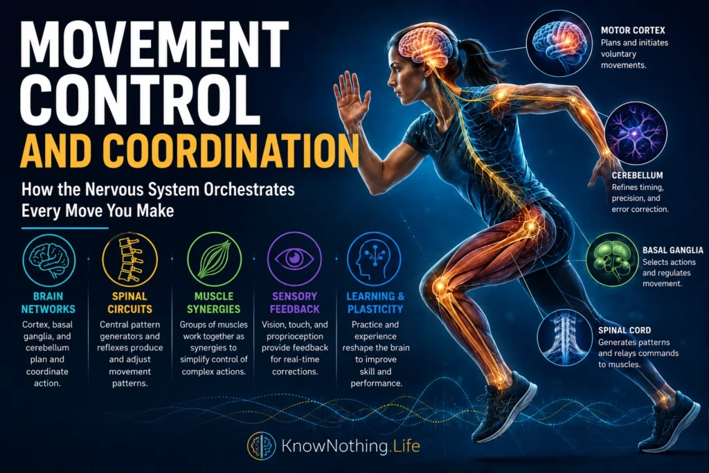
Movement control is the process through which the nervous system transforms intentions, sensory information, and bodily states into organized muscular activity. Coordination refers to the way muscles, joints, limbs, and neural systems cooperate so that movement remains accurate, stable, and appropriate to a goal. Even a simple reach requires the brain to estimate the hand’s current position, select a path, generate forces across several joints, compensate for gravity, and respond to unexpected resistance. Walking, speaking, writing, and playing an instrument require even more elaborate coordination across rapidly changing sequences.
No single brain structure contains a complete movement program. Voluntary action emerges through interactions among the cerebral cortex, basal ganglia, cerebellum, brainstem, spinal cord, peripheral nerves, muscles, and sensory receptors. These systems operate at different levels and timescales. The cortex contributes to planning and flexible control, the basal ganglia influence action selection, the cerebellum predicts consequences and corrects errors, and spinal circuits translate descending commands into coordinated patterns. Movement is therefore a distributed achievement in which prediction and feedback continuously shape one another.
Spinal Circuits and the Foundations of Movement
The spinal cord contains motor neurons that directly activate skeletal muscles, but it also contains interneuronal circuits capable of organizing complex patterns. Reflex pathways automatically adjust muscle activity when receptors detect changes in stretch, force, or contact. These responses help maintain posture and protect the body without requiring every correction to be consciously planned. Spinal networks can also generate alternating activity across groups of flexor and extensor muscles, providing a foundation for rhythmic behaviors such as walking and swimming.
Such networks are commonly called central pattern generators. In experiments on isolated lamprey spinal preparations, Sten Grillner, Alan McClellan, and Carl Perret found that mechanically bending part of the spinal cord could reset and synchronize the rhythmic motor pattern associated with swimming. The results showed that spinal circuits can generate a basic rhythm while remaining highly responsive to sensory information. Normal locomotion consequently depends neither on a completely prewritten spinal program nor on continuous control of every muscle from the brain. It emerges from interaction among central rhythms, descending goals, and feedback from the moving body.
Motor Cortex and Population Activity
The motor cortex contributes strongly to voluntary movement, particularly actions requiring flexible control of the hands, arms, face, and speech muscles. Its organization is often illustrated with a motor homunculus, but cortical control is not divided into perfectly isolated regions for individual muscles. Neural populations overlap, and one neuron may become active during several movements. The meaning of its activity depends on the wider network, the task, and the pattern evolving through time.
Apostolos Georgopoulos, Andrew Schwartz, and Ronald Kettner demonstrated this distributed organization in their 1986 study, Neuronal Population Coding of Movement Direction. Individual motor-cortical neurons responded broadly around preferred reaching directions, but combining the responses of many cells produced a population vector that closely followed the actual movement. Mark Churchland and colleagues later showed that population activity develops through orderly rotational dynamics during reaching. Together, these studies suggest that movement is generated by changing patterns across neural populations rather than by separate command cells specifying every muscular contraction.
Prediction, Feedback, and Internal Models
Sensory feedback is essential for movement control, but it arrives with unavoidable delays. The nervous system cannot wait for a completed movement before determining whether it is going wrong. It must estimate the body’s current state from vision, touch, proprioception, balance signals, and copies of outgoing motor commands. These estimates allow rapid corrections when an object moves, the ground becomes unstable, or a lifted object is heavier than expected.
David Wolpert, Zoubin Ghahramani, and Michael Jordan presented influential evidence for an internal model in their 1995 study, An Internal Model for Sensorimotor Integration. Their results supported the idea that the nervous system predicts the sensory consequences of movement and combines those predictions with actual feedback. Internal models help explain why familiar actions can remain smooth despite neural delays and why self-produced sensations are treated differently from external ones. Prediction is not a substitute for feedback; it provides a provisional estimate that incoming sensory evidence can confirm or correct.
The Cerebellum and Error-Based Coordination
The cerebellum is central to movement timing, accuracy, adaptation, and coordination. Damage to it usually does not cause complete paralysis. Instead, movements may become irregular, poorly timed, unstable, or unable to stop at the intended location. These symptoms indicate that the cerebellum does not simply initiate muscle activity. It helps compare intended actions with their actual or predicted consequences and uses mismatches to improve future commands.
Adaptation experiments demonstrate this function clearly. Reza Shadmehr and Ferdinando Mussa-Ivaldi asked participants to make arm movements while a robotic device produced unfamiliar forces. With practice, participants generated compensatory commands and later showed aftereffects when the force was removed, indicating that they had learned a model of the altered dynamics. Human prism-adaptation research has likewise found substantially reduced adaptation in people with cerebellar disorders. These findings connect the cerebellum with error-driven updating that allows movements to remain coordinated when bodies, tools, or environments change.
Basal Ganglia and Action Selection
The basal ganglia help determine which actions are initiated, suppressed, continued, or modified. They participate in loops connecting the cortex, striatum, pallidum, substantia nigra, and thalamus. These circuits are involved not only in movement onset but also in effort, speed, habit formation, reward-based learning, and the selection of one behavior among competing possibilities. Their importance becomes evident in Parkinson’s disease, where dopamine loss disrupts movement initiation and scaling, and in conditions involving unwanted or poorly controlled actions.
Older accounts often described the basal-ganglia direct pathway as promoting movement and the indirect pathway as suppressing it. Modern experiments reveal a more dynamic relationship. Hao Li and Xin Jin recorded and manipulated pathway-specific activity in mice performing an action-selection task. They found that direct-pathway effects were comparatively linear, while indirect-pathway effects depended nonlinearly on network state and incoming information. The study suggests that the basal ganglia coordinate choice through interacting signals that facilitate selected actions, regulate alternatives, and adapt decisions to context rather than operating as a simple accelerator and brake.
Muscle Synergies and the Degrees-of-Freedom Problem
The human body contains many joints and hundreds of muscles, creating an enormous number of possible combinations for accomplishing even one task. The nervous system does not appear to calculate each muscle independently on every occasion. One proposed solution is the use of muscle synergies: coordinated patterns in which groups of muscles are activated together as functional units. A smaller collection of reusable patterns could simplify control while still permitting flexible behavior.
Andrea d’Avella, Philippe Saltiel, and Emilio Bizzi examined muscle activity during natural frog movements and found that complex behaviors could be reconstructed by combining a limited number of time-varying synergies. Later work by d’Avella and Bizzi found both shared and movement-specific synergies across a wider range of behaviors. These studies do not prove that every coordinated action is stored as a fixed module, and some apparent synergies may partly reflect biomechanics or task constraints. They nevertheless show that structured combinations can reduce the dimensional complexity of muscular control.
Optimal Feedback and Flexible Coordination
Perfectly repeating the same joint trajectory is rarely necessary. When carrying a cup, the crucial goal may be to prevent spilling, while the elbow and shoulder can vary within limits. Optimal feedback control proposes that the nervous system corrects deviations that threaten the task while tolerating variability that does not matter. Coordination is therefore not the elimination of all variation. It is the intelligent regulation of task-relevant variation.
Emanuel Todorov and Michael Jordan developed this framework in their 2002 paper, Optimal Feedback Control as a Theory of Motor Coordination. Their model explained why the nervous system may allow several body parts to vary independently until their combined behavior threatens the goal. It also predicted rapid corrective responses and flexible muscle coordination without requiring one rigid trajectory. This view helps unite sensory feedback, motor redundancy, and goal-directed action: the nervous system continually chooses corrections according to their usefulness, energetic cost, uncertainty, and behavioral consequences.
Motor Learning, Plasticity, and Rehabilitation
Movement control changes through practice. Early attempts at a skill usually require attention and produce large errors, whereas repeated performance becomes faster, more stable, and less mentally demanding. Learning can update internal models, strengthen useful sensory predictions, refine muscle combinations, and reorganize cortical representations. It does not merely strengthen one fixed pathway; different stages of learning recruit changing combinations of cortical, cerebellar, basal-ganglia, and spinal mechanisms.
Randolph Nudo and colleagues trained monkeys to perform a skilled hand task and found use-dependent changes in the functional representations of primary motor cortex. Practiced movements gained cortical territory when training demanded genuine skill, supporting the idea that motor maps remain plastic in adulthood. Such findings have influenced rehabilitation after stroke and other neurological injuries. Repetitive practice is most effective when it is purposeful, challenging, and connected to meaningful sensory feedback, because recovery depends on teaching surviving networks how to coordinate action again rather than simply exercising muscles in isolation.
Movement control and coordination ultimately arise from cooperation across the nervous system. Spinal networks generate and adjust patterns, sensory pathways report the body’s state, cortical populations organize flexible commands, the cerebellum corrects errors, and the basal ganglia select actions according to goals and outcomes. Muscle synergies and feedback-control strategies help manage the body’s many degrees of freedom. The result is not mechanical perfection but adaptable stability: the capacity to reach the same goal through slightly different movements as the body and environment continually change.


