
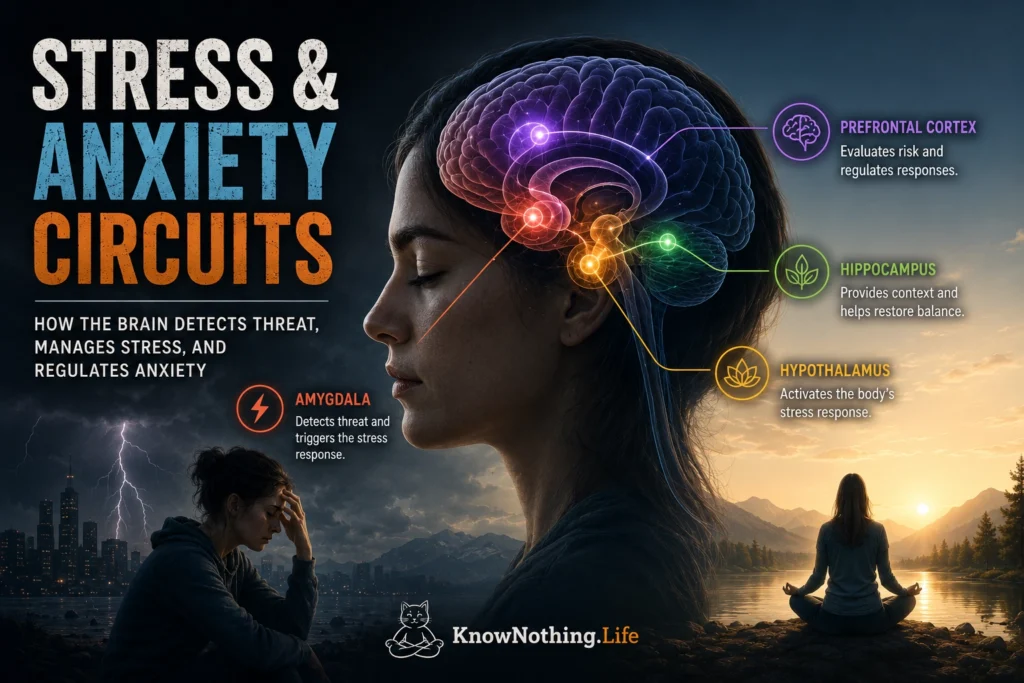
Stress and anxiety arise from coordinated brain and body systems that detect challenges, estimate danger, mobilize energy, and prepare adaptive behavior. Stress generally describes the response to a demand that is occurring or expected, while anxiety emphasizes anticipation, uncertainty, and the possibility of future harm. Both can be protective. A temporary stress response increases alertness and makes metabolic resources available, while anxiety encourages caution when outcomes are uncertain. Problems develop when these systems remain active without sufficient danger, respond too intensely, or fail to return to baseline after a challenge has passed.
There is no single stress center or anxiety center in the brain. These states emerge through communication among the amygdala, bed nucleus of the stria terminalis, hippocampus, hypothalamus, insula, anterior cingulate cortex, prefrontal cortex, locus coeruleus, brainstem, and endocrine system. Some pathways produce immediate defensive reactions, while others sustain vigilance, monitor the body, represent context, imagine future outcomes, or regulate emotional responses. Experiments contrasting predictable and unpredictable aversive events have found partly different patterns of neural activity during brief fear and sustained anxiety, supporting the view that these states overlap without being neurologically identical.
The HPA Axis and the Biological Stress Response
One of the central stress pathways is the hypothalamic-pituitary-adrenal axis, commonly called the HPA axis. When a challenge is evaluated as significant, neurons in the hypothalamus release corticotropin-releasing hormone. This stimulates the pituitary gland to secrete adrenocorticotropic hormone, which travels through the bloodstream to the adrenal glands and promotes cortisol release. Cortisol helps redistribute energy, alter immune activity, and support adaptation during demanding situations. At the same time, sympathetic nervous system activity can increase heart rate, breathing, blood pressure, sweating, and muscular readiness.
Clemens Kirschbaum and colleagues demonstrated these coordinated responses with the Trier Social Stress Test, in which participants prepare a speech and perform mental arithmetic before an evaluative audience. Across several experiments, the procedure reliably increased heart rate and produced substantial elevations in ACTH and cortisol. The study revealed that social evaluation, uncertainty, and loss of control can activate biological stress systems even in the absence of physical danger. Stress circuitry therefore responds not only to injury or direct threat but also to situations in which social standing, competence, or future outcomes appear uncertain.
Cortisol also participates in feedback loops that help terminate the response. Receptors in the hippocampus, prefrontal cortex, hypothalamus, and other regions detect circulating glucocorticoids and influence whether HPA activity continues. Tony Buchanan, Daniel Tranel, and Kirschbaum found that people with bilateral hippocampal damage did not show the typical cortisol increase during psychosocial stress, although their heart-rate and subjective emotional responses remained largely intact. The results indicate that the hippocampus contributes actively to organizing human endocrine stress responses rather than functioning only as a memory structure.
The Amygdala, Insula, and Threat Significance
The amygdala helps determine which events deserve immediate attention. Its nuclei receive sensory, contextual, and bodily information and help form associations between cues and aversive outcomes. The amygdala can influence the hypothalamus, brainstem, striatum, hippocampus, and cortex, allowing threat-related information to alter attention, memory, autonomic activity, and behavior. It is often described as a fear center, but its broader function is to identify biological significance. It responds to uncertainty, novelty, reward, social signals, and emotionally important events as well as obvious danger.
Stress can change the way the amygdala communicates with the rest of the brain. In a human resting-state imaging experiment, Inga Veer and colleagues found increased connectivity between the amygdala and cortical midline regions after acute psychological stress. Cortisol levels have also been associated with differences in amygdala connectivity with medial prefrontal regions, suggesting that endocrine and neural components of stress operate as an integrated system. These changes may temporarily prioritize emotional monitoring and internally focused processing after a stressful event.
The insular cortex contributes by representing the internal condition of the body. It processes signals related to heartbeat, breathing, pain, temperature, nausea, and muscular tension, helping the brain interpret whether bodily arousal indicates danger. Lisa Somerville and colleagues found that the anterior insula and bed nucleus of the stria terminalis tracked changing environmental threat levels during an uncertain-threat task. People with greater trait anxiety showed stronger hypervigilant monitoring, illustrating how attention to both external uncertainty and internal sensations can sustain an anxious state.
The BNST and Sustained Anxiety
The bed nucleus of the stria terminalis, or BNST, is part of the extended amygdala and is strongly connected with the amygdala, hypothalamus, hippocampus, ventral striatum, and brainstem. Although the amygdala is often emphasized during rapid responses to clear cues, the BNST is especially important when danger is uncertain, diffuse, or difficult to predict. Anxiety frequently occurs in precisely these conditions: a person knows that something unpleasant might happen but does not know when, where, or how severe it will be.
In human imaging research, Somerville and colleagues found sustained BNST activity as participants monitored fluctuating environmental danger. Robert Alvarez and colleagues similarly reported distinct neural patterns during brief, predictable threat and prolonged, unpredictable threat. The findings do not establish a perfect division in which the amygdala produces fear and the BNST produces anxiety, because both regions participate in several defensive processes. They instead suggest that the BNST is particularly relevant when the nervous system must maintain vigilance over time.
Sung-Yon Kim and colleagues demonstrated the complexity of BNST function by manipulating specific neural projections in mice. Different output pathways controlled separable features of anxiety-related behavior, including avoidance, breathing changes, and risk assessment. Rather than generating one unified anxiety response, the BNST helped assemble a larger defensive state from multiple components. Chronic stress can also alter BNST structure and signaling. Ajai Vyas and colleagues observed increased dendritic arborization in BNST neurons following chronic immobilization stress, providing a possible cellular mechanism through which repeated stress may strengthen sustained anxiety.
The Hippocampus and the Context of Danger
The hippocampus helps the brain determine where and when danger occurs. It represents environments, relationships among events, and the contextual details surrounding an experience. This information allows a defensive response to remain tied to the situation in which it was learned. Without accurate contextual processing, fear and anxiety may generalize beyond the original danger, causing safe locations or harmless sensations to acquire threatening meaning.
Human imaging research has found that anterior and posterior hippocampal regions relate differently to state and trait anxiety. An experiment by Ajay Satpute and colleagues associated anterior hippocampal activity with momentary anxiety and posterior hippocampal characteristics with more stable anxiety tendencies. Other studies of contextual fear have found coordinated involvement of the hippocampus, amygdala, and ventromedial prefrontal cortex when people retrieve either danger or extinction memories. These systems help decide whether an old defensive response remains appropriate in the present setting.
Stress hormones can change hippocampal memory processing. Juraj Kukolja and colleagues administered hydrocortisone, a norepinephrine reuptake inhibitor, or both to healthy participants during emotional memory encoding. Cortisol alone reduced hippocampal responses to emotional material, whereas combined elevation of cortisol and norepinephrine enhanced hippocampal activity. The study demonstrates that stress effects cannot be attributed to cortisol in isolation. Their consequences depend on timing, arousal, neurotransmitter interactions, and the type of information being processed.
The Locus Coeruleus and Hypervigilance
The locus coeruleus is a small brainstem structure that supplies much of the brain’s norepinephrine. When a stressor appears, locus-coeruleus activity can increase alertness, orient attention toward important information, and amplify sensory processing. Moderate activation supports flexible attention and rapid adaptation. Excessive or prolonged activation can contribute to distractibility, sleep disruption, exaggerated startle, racing thoughts, and hypervigilance.
Tae-Ho Lee and colleagues found that arousal increased neural gain through mechanisms associated with the locus coeruleus–norepinephrine system in younger adults. Neural gain refers to the strengthening of responses to highly prioritized information while weaker signals receive less processing. During stress, this can make a possible threat unusually prominent and difficult to ignore. The same mechanism may be helpful in an emergency yet counterproductive when neutral events are repeatedly treated as urgent.
Norepinephrine also affects the extended amygdala. Experiments by Marco Cecchi and colleagues showed that stress-induced norepinephrine activity in the lateral BNST facilitated anxiety-like behavior and influenced HPA-axis activation in rats. The result illustrates how brainstem arousal systems, sustained-threat circuits, and hormonal responses reinforce one another. Anxiety is therefore not merely a pattern of worried thought; it is a coordinated state involving attention, bodily activation, memory, and defensive motivation.
The Prefrontal Cortex and Emotional Regulation
The prefrontal cortex helps evaluate whether a threat is real, compare short- and long-term consequences, maintain goals, reinterpret situations, and suppress responses that no longer fit the evidence. Ventromedial prefrontal regions participate in safety learning and the regulation of amygdala-centered responses, while dorsolateral regions support working memory and deliberate control. These systems make it possible to remain flexible rather than reacting automatically to every sign of uncertainty.
Acute stress can temporarily weaken this control. Shaozheng Qin and colleagues exposed healthy participants to psychological stress before a working-memory task and found reduced activity in the dorsolateral prefrontal cortex. When prefrontal processing becomes less efficient, attention may narrow, habitual responses may dominate, and it can become more difficult to evaluate complex situations objectively. This helps explain why people under intense pressure may know that a worry is exaggerated yet struggle to disengage from it.
Repeated stress can produce longer-lasting plasticity. Susan Cook and Cara Wellman found reduced length and branching of apical dendrites in the medial prefrontal cortex of chronically stressed rats. In contrast, Vyas and colleagues observed dendritic expansion in parts of the basolateral amygdala alongside atrophy in hippocampal neurons. These opposing changes suggest that chronic stress may strengthen systems involved in emotional vigilance while weakening networks responsible for context, memory, and regulation. Importantly, Jason Radley and colleagues later showed that some prefrontal dendritic changes reversed after a recovery period, demonstrating that stress-related neural remodeling is not necessarily permanent.
When Adaptive Stress Becomes Chronic Anxiety
Healthy stress circuits activate when needed and gradually settle when safety returns. Chronic anxiety can develop when uncertain situations repeatedly recruit threat monitoring, when bodily sensations are interpreted catastrophically, when danger memories generalize, or when regulatory systems fail to retrieve evidence of safety. Anxiety disorders therefore cannot be reduced to one overactive region. They involve changing interactions among the amygdala, BNST, insula, hippocampus, prefrontal cortex, locus coeruleus, autonomic nervous system, and HPA axis.
Stress and anxiety circuits also remain capable of learning. Safety experiences, cognitive reappraisal, exposure-based therapy, sleep, exercise, supportive relationships, and predictable environments can provide new information that changes how threat is evaluated. The aim is not to eliminate the stress response, which remains essential for survival, but to restore flexibility. An adaptive nervous system can mobilize rapidly during genuine danger, distinguish uncertainty from catastrophe, and return the body and mind to a state in which attention can once again be directed toward exploration, connection, and long-term goals.


