
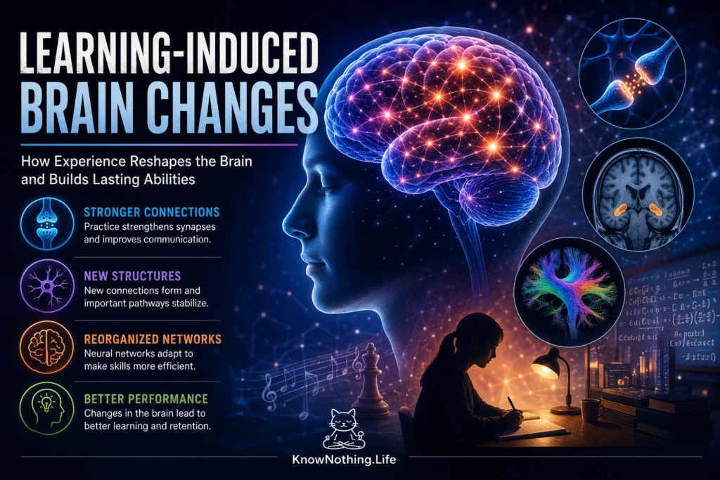
Learning is not merely the collection of information. It is a biological process that alters how the nervous system responds to future events. When a person practices a movement, memorizes a route, studies a language, or learns to distinguish unfamiliar sounds, repeated activity changes the neural circuits involved in that task. Some changes occur rapidly through adjustments in synaptic strength, while others develop over days, months, or years through changes in dendritic spines, cortical organization, white matter, and patterns of communication between brain regions.
These learning-induced changes are commonly described as neuroplasticity, but the term covers several distinct processes. Learning may strengthen selected connections while weakening competing ones, increase the efficiency of a network, alter the excitability of neurons, or change which brain regions are recruited during performance. Structural brain changes detected with magnetic resonance imaging do not necessarily represent the growth of entirely new tissue. They may reflect combinations of altered synapses, blood vessels, glial cells, dendrites, axons, or myelin. Learning reshapes the brain through coordinated changes at several biological levels rather than through one universal mechanism.
Synaptic Plasticity and the Formation of Memory
At the cellular level, learning depends heavily on synaptic plasticity—the ability of connections between neurons to become stronger or weaker. Long-term potentiation increases synaptic responsiveness after coordinated activity, while long-term depression reduces the influence of selected connections. These opposing processes allow neural networks to emphasize associations that repeatedly predict important outcomes and weaken relationships that are inaccurate or no longer useful. Changes in glutamate receptors, calcium signaling, protein phosphorylation, gene expression, and local protein synthesis can help convert brief patterns of activity into more persistent alterations in synaptic function.
Jonathan Whitlock and colleagues provided direct evidence connecting behavioral learning with synaptic potentiation in their 2006 study Learning Induces Long-Term Potentiation in the Hippocampus. Rats that experienced one-trial inhibitory avoidance learning showed hippocampal changes resembling experimentally induced LTP, including related modifications in glutamate receptors. Learning also reduced the amount of additional potentiation that researchers could later produce in the affected pathway, suggesting that the behavioral experience and laboratory stimulation had recruited overlapping synaptic mechanisms. The study supported the view that learning leaves measurable physiological traces within the circuits used to form memories.
Learning Builds and Stabilizes Synaptic Connections
Learning can also change the physical structure of synapses. Dendritic spines are small protrusions that receive many excitatory inputs, and their formation, enlargement, shrinkage, or disappearance can alter how neurons participate in a circuit. These structures are dynamic, particularly during development, but the adult brain retains the ability to remodel them. Learning does not simply produce unlimited numbers of new connections. Instead, experience appears to encourage the formation of candidate synapses, after which some are stabilized and others are removed.
In 2009, Tianyi Xu and colleagues used repeated imaging to observe individual dendritic spines in the motor cortex of living mice learning a forelimb-reaching task. Their study, Rapid Formation and Selective Stabilization of Synapses for Enduring Motor Memories, found that training stimulated rapid spine formation, with some new spines appearing within an hour. Although overall spine density later moved back toward its earlier level as other spines disappeared, a selected proportion of the learning-associated spines remained stable. Different motor experiences produced partly distinct patterns of spine formation, suggesting that enduring skills may be supported by the selective stabilization of particular synaptic connections.
Practice Reorganizes Functional Brain Activity
Learning changes not only individual synapses but also the activity of larger neural systems. When a skill is new, performance may require substantial attention and widespread recruitment of cortical areas. As practice continues, activation patterns can become more specialized, stable, or efficient. The brain may rely less heavily on conscious control and more on task-specific networks that have been refined by repetition. This is one reason a practiced movement can eventually feel automatic even though it initially required deliberate concentration.
Avi Karni and colleagues demonstrated experience-dependent change in the adult motor cortex using functional magnetic resonance imaging. In their 1995 study Functional MRI Evidence for Adult Motor Cortex Plasticity During Motor Skill Learning, participants practiced a sequence of finger movements over several weeks. Improvements in performance were accompanied by changes in the representation of the trained sequence within the primary motor cortex. Later research by Karni and colleagues distinguished fast improvements occurring during initial practice from slower changes developing between sessions and across days. These findings helped establish that skill learning unfolds through multiple phases supported by evolving patterns of neural activity.
Learning Can Alter Gray Matter
Human brain-imaging studies have reported measurable structural changes following periods of training. One of the best-known examples came from Bogdan Draganski and colleagues, who examined adults learning to juggle. In the 2004 study Neuroplasticity: Changes in Grey Matter Induced by Training, participants who acquired juggling skills showed temporary gray-matter increases in regions involved in processing visual motion. Some of these changes diminished after participants stopped practicing, indicating that learning-related anatomy can be dynamic rather than permanently fixed.
Training-related changes can also involve connected regions rather than one isolated cortical area. Martin Taubert and colleagues studied participants learning a whole-body balancing task and reported changes in cortical areas associated with movement as well as in nearby fiber pathways. Performance improvement was related to a complex pattern of structural increases and decreases over the training period. These findings caution against interpreting every local increase as improvement or every decrease as loss. Learning may involve expansion, refinement, redistribution, and increased efficiency, depending on the brain region and stage of training.
White Matter and Myelin Also Respond to Learning
The effects of learning extend beyond gray matter. White matter contains axons connecting distant parts of the nervous system, many of which are wrapped in myelin produced by oligodendrocytes. Myelin supports rapid and precisely timed neural transmission. Because complex behavior depends on signals arriving across distributed networks at appropriate times, changes in white-matter organization may influence how efficiently brain regions communicate during a learned task.
In 2009, Jan Scholz and colleagues trained adults to juggle and found changes in diffusion-based measurements of white-matter architecture. Their study provided early evidence that white matter in the healthy adult brain can change in association with learning. Animal experiments later offered stronger evidence that active myelination contributes directly to skill acquisition. Ian McKenzie and colleagues found that preventing the formation of new oligodendrocytes impaired mice learning to run on a complex wheel. Lin Xiao and colleagues subsequently reported that rapid production of new oligodendrocytes was required during both early and later stages of motor-skill learning. Together, these studies suggest that learning modifies not only synaptic connections but also the biological systems that coordinate communication between them.
Long-Term Expertise and the Hippocampus
Learning-related brain differences can accumulate over years when a task places sustained demands on a particular cognitive system. Eleanor Maguire and colleagues studied licensed London taxi drivers, who traditionally learned a detailed mental representation of thousands of streets and landmarks. Their 2000 study found that taxi drivers had greater relative gray-matter volume in posterior portions of the hippocampus than control participants. Posterior hippocampal volume was also related to the amount of time spent working as a taxi driver, although the original cross-sectional study could not by itself establish causation.
Later research strengthened the link between navigation learning and hippocampal change. Taxi drivers differed from London bus drivers, whose driving experience and occupational stress were comparable but whose routes were more restricted. Most importantly, Katherine Woollett and Maguire followed people while they trained to acquire London’s navigational qualification. Those who successfully learned the city’s layout showed increased posterior hippocampal gray matter over the multiyear training period, while unsuccessful trainees and control participants did not show the same pattern. The findings provided longitudinal evidence that intensive real-world learning can contribute to regionally specific structural change.
Learning Changes Networks Selectively
Brain change is not proportional to repetition alone. Learning is influenced by attention, error correction, motivation, novelty, difficulty, reward, and the relevance of feedback. Repeating a fully mastered action may produce less reorganization than practicing a demanding task that requires continual adjustment. Different stages of learning may also recruit different mechanisms. Early improvement can depend on strategy and rapid synaptic change, while long-term retention may require protein synthesis, structural stabilization, altered network coordination, and continued practice.
The changes are also selective. Learning one skill does not make the entire brain uniformly larger or more efficient. It modifies the pathways most involved in processing the task, often alongside systems that support attention, error monitoring, memory, and action selection. Improvements may therefore transfer poorly to unrelated abilities. A trained brain is not simply a stronger brain; it is a brain increasingly organized around the demands it has repeatedly encountered.
The Limits of Interpreting Brain Change
Learning-related changes detected in imaging studies must be interpreted carefully. Functional MRI measures changes associated with blood oxygenation rather than neuronal firing directly, while structural MRI cannot usually identify the exact cellular process behind an apparent gray-matter difference. Diffusion imaging provides information related to white-matter microstructure but does not directly photograph newly produced myelin. Researchers must therefore combine human imaging with behavioral experiments, animal models, electrophysiology, microscopy, and molecular methods.
Not every brain change is beneficial, permanent, or exclusive to learning. Fatigue, stress, sleep, age, prior experience, and measurement variability can influence results. Some learning-induced changes decline when practice stops, while others remain stable for years. Plasticity can also support maladaptive learning, including persistent fear, addiction, compulsive behavior, and chronic pain. The nervous system adapts to repeated experience according to biological significance and activity patterns, not according to whether a learned response is healthy.
Why Learning-Induced Brain Changes Matter
Research on learning-induced brain change has replaced the older view of the adult brain as a largely fixed organ. Experience can modify synapses within minutes, stabilize new dendritic spines over days, reorganize functional networks with practice, and alter measurable gray- and white-matter properties over longer periods. These processes allow the nervous system to preserve useful information while remaining capable of revision.
The evidence also gives learning a physical dimension. Knowledge and skill are not stored in one location or represented by one kind of change. They emerge from modified patterns of connection, timing, structure, and activity distributed across neural systems. Learning changes the brain because the ability to behave differently in the future requires the nervous system itself to become different. The brain retains continuity not by remaining unchanged, but by continually reorganizing experience into more durable patterns.


