
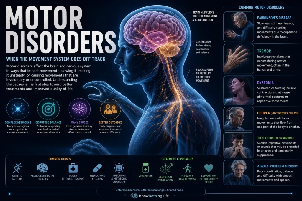
Motor disorders are conditions that interfere with the planning, initiation, timing, force, coordination, or execution of movement. They can produce actions that are too slow, too small, excessive, repetitive, unstable, weak, or poorly coordinated. Some primarily disrupt basal-ganglia circuits, as in Parkinson’s disease, dystonia, Huntington’s disease, and tic disorders. Others arise from cerebellar degeneration or damage affecting the motor cortex, corticospinal tracts, spinal cord, peripheral nerves, neuromuscular junctions, or muscles. The term therefore describes a broad clinical problem rather than one specific diagnosis. A person may understand exactly what action is intended while the nervous system fails to select the correct movement, suppress unwanted activity, generate sufficient force, or correct an error.
Neurologists often distinguish movement disorders from disorders of strength. Movement disorders typically produce involuntary activity or impaired regulation despite relatively preserved basic muscular power. Damage to corticospinal pathways, motor neurons, peripheral nerves, or muscles is more likely to cause weakness, paralysis, spasticity, or abnormal fatigue. These categories still overlap because every voluntary action depends on communication among the cortex, basal ganglia, cerebellum, brainstem, spinal cord, nerves, and muscles. Diagnosis begins by identifying the movement pattern—such as tremor, rigidity, bradykinesia, dystonia, chorea, tic, myoclonus, or ataxia—and then determining which network, disease process, medication, toxin, or injury could have produced it.
Parkinsonism and Hypokinetic Movement
Parkinsonism is characterized by bradykinesia, or slowness accompanied by a progressive reduction in movement, together with features such as rigidity, resting tremor, or postural instability. Parkinson’s disease is its best-known cause, although medications that block dopamine receptors, vascular damage, and several neurodegenerative diseases can produce related syndromes. The biological foundation of Parkinson’s disease includes degeneration of dopamine-producing neurons and severe loss of dopamine within the striatum. Early work by Oleh Hornykiewicz connected striatal dopamine deficiency with Parkinsonian symptoms. Maria Spillantini and colleagues later demonstrated that alpha-synuclein is a major component of Lewy bodies, one of the characteristic pathological findings associated with the disease.
Loss of dopamine disrupts basal-ganglia circuits involved in initiating actions, scaling movement, forming habits, and regulating effort. Levodopa can restore part of the missing dopamine signal and improve slowness and rigidity, although prolonged treatment may be accompanied by fluctuations and involuntary dyskinesias. For carefully selected people with advanced disease, deep brain stimulation can modify pathological network activity. In a randomized trial led by Günther Deuschl, subthalamic stimulation combined with medication produced greater improvements in motor function and quality of life than medical management alone, although surgery and stimulation also introduced important risks.
Tremor and Dystonia
Tremor is a rhythmic oscillation created by recurring activity within motor networks. Essential tremor commonly appears while maintaining posture or performing an action, especially with the hands and arms, although the head and voice may also be affected. Its biology cannot be reduced to one damaged nucleus. In a positron-emission tomography study, Ian Jenkins and colleagues found increased bilateral cerebellar activity in people with essential tremor both at rest and during tremor. Later network mapping connected abnormalities reported throughout the brain to a shared system involving the cerebellum, thalamus, motor cortex, and related regions, with the cerebellum functioning as a central hub.
Dystonia produces sustained or intermittent muscle contractions that cause twisting movements, repetitive actions, or abnormal postures. It may affect one body region, such as the neck, eyelids, or hand, or become more generalized. Research increasingly describes dystonia as a disorder of distributed sensorimotor control involving abnormal inhibition, sensory processing, plasticity, and communication among basal-ganglia, cerebellar, and cortical networks. Alessandra Quartarone and colleagues found unusually broad sensorimotor plasticity in people with focal hand dystonia, including abnormalities beyond the symptomatic body part. Controlled trials have shown that botulinum toxin can reduce abnormal muscular activity in cervical dystonia, while pallidal stimulation can benefit some severe, treatment-resistant cases.
Chorea, Tics, and Other Hyperkinetic Disorders
Chorea consists of irregular, unpredictable movements that appear to flow from one body part to another. Huntington’s disease is a major inherited cause and combines motor symptoms with changes in cognition, behavior, and emotional regulation. In 1993, the Huntington’s Disease Collaborative Research Group identified an expanded CAG repeat in the huntingtin gene, establishing the mutation responsible for the disorder. Neuropathological studies later demonstrated selective vulnerability among striatal projection systems, with some neuronal pathways deteriorating earlier than others. Early disruption of circuits that normally suppress competing movements can contribute to chorea, while more extensive degeneration eventually produces rigidity, slowness, impaired balance, and loss of voluntary control.
Tics are brief movements or sounds that may be temporarily suppressed and are often preceded by a compelling bodily urge. Tourette syndrome is characterized by recurring motor and vocal tics that typically begin during development. Brain imaging does not support the existence of one isolated “tic center.” In a functional MRI study, Tara Wang and colleagues found excessive activity throughout motor portions of cortico-striato-thalamo-cortical circuits during spontaneous tics, accompanied by weaker activity in control-related regions such as the caudate and anterior cingulate cortex. Hyperkinetic disorders also include myoclonus, in which sudden shock-like movements may originate from cortical, subcortical, brainstem, spinal, or peripheral mechanisms. Similar-looking movements can therefore reflect very different neural abnormalities.
Ataxia and Cerebellar Dysfunction
Ataxia refers to impaired coordination rather than a simple loss of strength. It may produce a wide-based and unstable gait, inaccurate reaching, slurred speech, abnormal eye movements, or difficulty coordinating several joints. Ataxia can result from stroke, tumors, immune disease, toxins, nutritional deficiencies, inherited spinocerebellar conditions, or progressive cerebellar degeneration. Because the cerebellum helps predict sensory consequences and update motor commands from error, its damage makes movement more variable and reduces the nervous system’s ability to adapt, even when the muscles remain capable of generating force.
Ya-Weng Tseng and colleagues examined people with hereditary cerebellar ataxia during visually perturbed reaching. Participants could observe their errors but adapted much less than healthy controls, whether or not they were able to make immediate corrections during the movement. The results showed that cerebellum-dependent learning relies strongly on sensory prediction errors—the differences between the expected and observed consequences of a motor command. Later research similarly found impaired feedforward adaptation across reaching and speech among people with cerebellar degeneration, even when rapid feedback corrections remained comparatively preserved.
Diagnosis and Treatment
Motor disorders are diagnosed first through careful observation. Clinicians examine whether an abnormal movement appears at rest, during posture, or during voluntary action; whether it is rhythmic or irregular; whether it can be suppressed; and whether weakness, sensory loss, cognitive change, medication exposure, or a family history is present. Blood tests, genetic analysis, electromyography, nerve-conduction studies, MRI, metabolic imaging, and dopamine-system imaging may answer particular questions, but no single test identifies every disorder. The same symptom can have several causes, while one disease may produce different movement patterns as it progresses.
Treatment varies because abnormal movement may result from deficient neurotransmission, excessive neural activity, lost neurons, maladaptive plasticity, or structural damage. Levodopa can improve dopamine-dependent symptoms in Parkinson’s disease, while propranolol can reduce essential tremor in some patients. Botulinum toxin weakens selected overactive muscles, and deep brain stimulation can modulate networks involved in Parkinsonism, tremor, or dystonia. Physical, occupational, and speech therapies address balance, dexterity, communication, and daily independence. These approaches frequently manage symptoms rather than reverse the underlying disease, but they demonstrate that movement can be improved by intervening at several levels of the motor system.
Motor disorders reveal that movement is not produced by muscles alone. Every action depends on distributed systems that must choose among possibilities, estimate bodily position, predict consequences, suppress interference, and learn from results. When one part of this network fails, the consequences may appear as slowness, tremor, twisting, uncontrolled movement, weakness, or poor coordination. Understanding the pattern provides the first clue to understanding the disrupted neural mechanism.


