
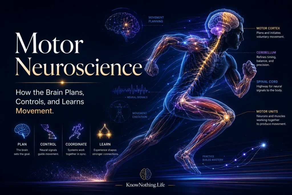
Motor neuroscience examines how the brain, spinal cord, peripheral nerves, muscles, and sensory systems work together to produce movement. Its subject matter ranges from basic reflexes and rhythmic activities such as walking to highly skilled actions such as speaking, writing, playing an instrument, or reaching for a fragile object. Movement may appear to begin when muscles contract, but successful action requires the nervous system to select a goal, estimate the body’s current state, create an appropriate command, predict its consequences, monitor the result, and correct errors. Motor control is therefore not a one-way transmission from the brain to the muscles. It is a continuous process of prediction, sensory feedback, and adjustment.
No single motor center controls the body independently. The spinal cord organizes reflexes and recurring movement patterns, the brainstem regulates posture and orientation, the motor cortex contributes to voluntary actions, the basal ganglia help select and initiate behavior, and the cerebellum refines timing and corrects errors. Parietal and premotor areas transform sensory information into possible actions, while prefrontal and limbic systems influence movement according to goals, motivation, and expected rewards. Motor neuroscience consequently studies distributed networks whose contributions overlap and change depending on whether an action is automatic, learned, externally triggered, or deliberately chosen.
The Spinal Cord, Motor Neurons, and Movement Patterns
Skeletal muscles are activated by lower motor neurons located in the spinal cord and brainstem. Each motor neuron and the muscle fibers it controls form a motor unit. Small motor units allow fine control of the fingers, face, and eyes, while larger units can generate the force needed for posture and locomotion. The nervous system varies muscular force by changing the firing rates of active motor neurons and recruiting additional units. Spinal interneurons coordinate these outputs with sensory information from the skin, joints, muscles, and tendons, allowing the body to respond rapidly to changes in load, position, and contact.
The spinal cord can also generate organized rhythmic activity through networks known as central pattern generators. These circuits can produce alternating patterns resembling swimming or walking without requiring a separate cortical instruction for every muscular contraction. Experiments on isolated lamprey spinal cords showed that spinal networks could generate locomotor rhythms and that movement-related sensory signals could reset or entrain those rhythms. Such findings do not mean that normal walking occurs without the brain or sensory feedback. Descending commands establish goals and speed, while sensory input continuously adjusts the basic pattern to terrain, balance, and unexpected obstacles.
Primary Motor Cortex and the Body Map
Primary motor cortex lies along the precentral gyrus of the frontal lobe and provides an important source of descending commands to the brainstem and spinal cord. Wilder Penfield and Edwin Boldrey’s 1937 electrical-stimulation studies helped establish that movements of different body parts can be evoked from different cortical regions. Their resulting motor map was often illustrated as a distorted human figure called the motor homunculus, in which the hands, face, and speech-related structures occupy unusually large territories. The enlargement reflects the complexity of their control rather than their physical size.
The map is useful but should not be interpreted as a collection of perfectly separated muscle switches. Edward Evarts found that the activity of pyramidal-tract neurons varied with the force exerted during voluntary movement, demonstrating that motor-cortical discharge is related to quantitative features of action. Later stimulation experiments by Michael Graziano and colleagues produced coordinated behaviors involving several joints, such as reaching, defensive movements, and bringing the hand toward the mouth. These results support a more flexible view in which motor cortex contains overlapping representations of muscles, movement parameters, and behaviorally useful actions.
Population Coding and Motor-Cortical Dynamics
Individual motor-cortical neurons usually respond during many different movements rather than representing only one muscle or direction. Apostolos Georgopoulos and colleagues found that many neurons are broadly tuned to preferred movement directions. The direction of a reach could be predicted more accurately by combining the activity of a population than by examining one cell alone. Their population-vector approach became an influential example of distributed neural coding, showing how precise behavior can emerge from many individually imprecise neurons.
More recent work has emphasized that motor cortex may operate as a dynamical system. Instead of simply representing static variables such as direction or force, a neural population may move through an evolving pattern of activity that generates the changing muscle commands needed for action. Mark Churchland and colleagues found strong rotational dynamics in motor-cortical populations during reaching, with preparatory activity establishing an initial neural state from which movement-related patterns unfolded. This perspective does not eliminate representations of direction, force, or muscles. It suggests that these properties may arise from the coordinated evolution of activity across an interconnected network.
Planning, Prediction, and Sensory Feedback
Voluntary movement is prepared before muscles begin to contract. Premotor cortex contributes strongly when actions are guided by external information, while supplementary motor areas are associated with internally generated movements, sequences, and coordination between the hands. Functional imaging of self-paced finger movement found supplementary motor area activation preceding activity in primary motor cortex. Preparatory signals do not necessarily encode a finished command waiting to be released. They may place the motor network into a state from which the intended movement can be produced efficiently.
Movement must also continue despite delayed and noisy sensory feedback. The nervous system appears to use internal models that predict how the body will respond to a command and what sensory consequences should follow. A forward model can estimate the next bodily state before feedback arrives, while comparison with actual feedback reveals an error that can guide correction. David Wolpert, Zoubin Ghahramani, and Michael Jordan provided behavioral evidence for an internal model in sensorimotor integration, and later computational work connected predictive control with the cerebellum, parietal cortex, and motor learning. These mechanisms allow a person to maintain stability, distinguish self-produced sensations, and adjust movement while it is still unfolding.
Basal Ganglia and the Selection of Action
The basal ganglia are interconnected subcortical nuclei that influence movement through loops connecting the cortex, striatum, pallidum, substantia nigra, and thalamus. They are involved in initiating actions, suppressing competing behaviors, regulating movement vigor, and learning from rewards. The influential model developed by Roger Albin, Anne Young, and John Penney described direct and indirect pathways with opposing influences on basal-ganglia output. Reduced dopamine in Parkinson’s disease was proposed to shift this balance toward excessive inhibition of thalamocortical movement systems, contributing to slowness, rigidity, and difficulty initiating action.
The classical pathway model remains useful, but modern findings show that basal-ganglia activity is more complex than a simple accelerator-and-brake arrangement. Direct and indirect pathway neurons can become active together during movement initiation, and their effects vary across cell populations and behavioral conditions. Experiments using selective optogenetic stimulation found that direct-pathway activity could facilitate movement by inhibiting particular basal-ganglia output neurons, whereas indirect-pathway activity could suppress movement by exciting other output neurons. The basal ganglia therefore help regulate which actions gain access to motor systems, how strongly they are performed, and whether their outcomes make them more likely to be repeated.
The Cerebellum and Motor Learning
The cerebellum contains more neurons than the rest of the brain combined and receives extensive information about motor commands, body position, and sensory consequences. Damage to it does not usually cause paralysis, but it can produce poorly timed, inaccurate, and unstable movement. The cerebellum contributes to coordination by predicting the consequences of commands and using errors to update future behavior. This is especially clear during adaptation tasks in which prisms, force fields, or altered tools create a mismatch between an intended action and its actual result.
Hiroshi Imamizu and colleagues used brain imaging to investigate people learning to control an unfamiliar computer mouse with a rotated relationship between hand and cursor movement. As learning progressed, activity within a localized part of the cerebellum reflected the acquired internal model of the new tool. Other theoretical accounts propose complementary learning roles for major motor systems: the cerebellum may specialize in error-based supervised learning, the basal ganglia in reward-based reinforcement learning, and cerebral cortex in acquiring flexible representations and skills. These divisions are not absolute, but they help explain why different forms of motor learning depend on interacting neural circuits.
Motor Disorders, Rehabilitation, and Neural Interfaces
Movement disorders reveal what different motor circuits contribute. Stroke can interrupt cortical or descending pathways, Parkinson’s disease disrupts basal-ganglia loops, cerebellar damage causes ataxia, and spinal cord injury separates supraspinal commands from lower motor circuits. Recovery depends partly on neural plasticity. Surviving networks can change their connectivity, sensory feedback can be retrained, and even spinal reflex pathways can undergo lasting modification through practice. Recent research showing structural changes in spinal neurons after reflex conditioning reinforces the view that motor learning is distributed across the nervous system rather than restricted to the cerebral cortex.
Brain–computer interfaces demonstrate how recorded motor signals can be translated into external action. Leigh Hochberg and colleagues showed that people with long-standing tetraplegia could use activity recorded from motor cortex to control a robotic arm, reach toward objects, and perform grasping actions. Such systems remain technically demanding and do not yet reproduce the speed, sensation, and adaptability of natural movement, but they reveal that intended actions can remain represented in motor networks after paralysis. The continuing goal of motor neuroscience is not merely to locate movement within the brain, but to explain how intention, prediction, sensation, learning, and muscular mechanics become coordinated behavior.


