
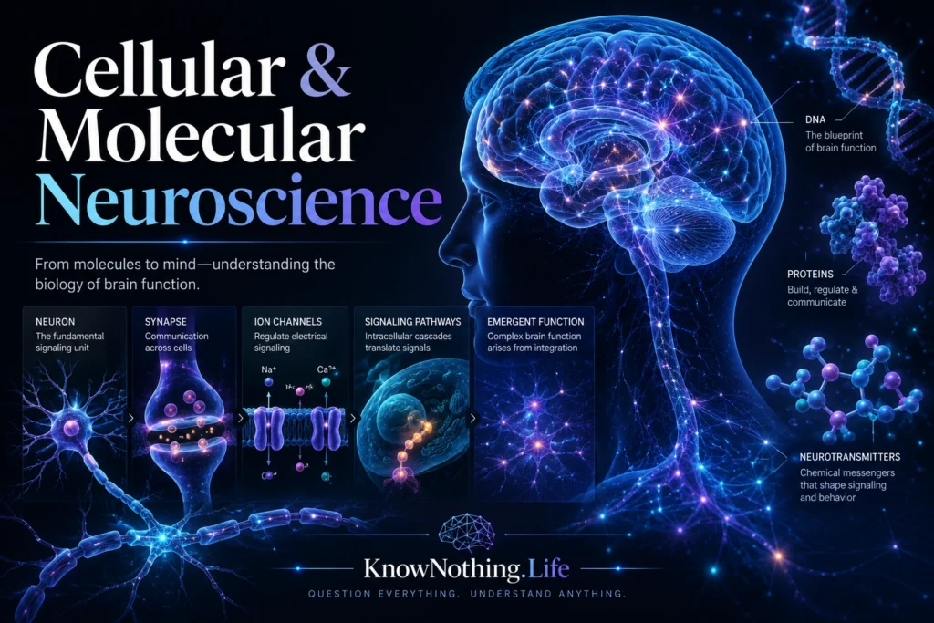
Cellular and molecular neuroscience is the study of how the nervous system works at the level of cells, molecules, membranes, genes, proteins, and synapses. While systems neuroscience looks at large networks and cognitive neuroscience studies functions such as memory, attention, perception, and emotion, cellular and molecular neuroscience asks what those functions are made of at the smallest biological scales. It examines neurons, glial cells, ion channels, neurotransmitters, receptors, synaptic vesicles, intracellular signaling pathways, gene expression, myelin, and cellular plasticity. In simple terms, it explains how the brain’s living cells generate electrical signals, communicate chemically, change with experience, and organize themselves into functional circuits.
This field matters because every large-scale brain function depends on small-scale biological mechanisms. A memory is not only a psychological event; it depends on synaptic changes, receptor trafficking, gene expression, and circuit reorganization. A movement is not only a command from the motor cortex; it depends on action potentials, neurotransmitter release, muscle activation, and feedback. A mood is not merely a feeling; it is shaped by neurotransmitter systems, hormones, receptor sensitivity, stress biology, immune signaling, and brain-body communication. Cellular and molecular neuroscience provides the foundation beneath all other branches of brain science.
Neurons as Living Signaling Cells
Neurons are specialized cells built to receive, process, and transmit information. Their structure reflects their function. Dendrites receive input from other cells, the soma supports metabolism and integrates signals, the axon carries electrical impulses, and synaptic terminals release chemical messengers. Neurons are electrically excitable because they maintain ion gradients across their membranes. When the right conditions are met, they can generate action potentials that travel rapidly along axons. NCBI’s neuroscience resources describe chemical synapses as specialized junctions in which synaptic vesicles in the presynaptic terminal release transmitter across the synaptic cleft.
The cellular level also reveals that neurons are not passive wires. They are living biochemical systems. They use mitochondria to generate energy, transport proteins and vesicles along axons, alter receptor expression, grow or retract synapses, and respond to injury or stimulation. Their ability to change is one of the reasons the brain can learn. A neuron is both a cell and a signal-processing unit, using membranes, ions, proteins, and molecular cascades to participate in perception, action, memory, and thought.
Ion Channels and Electrical Signaling
Ion channels are among the most important molecules in neuroscience. These protein pores allow charged particles such as sodium, potassium, calcium, and chloride to cross the cell membrane. Their opening and closing control membrane voltage, action potentials, synaptic responses, and neuronal excitability. The modern history of ion-channel physiology is closely tied to Alan Hodgkin and Andrew Huxley’s work on the squid giant axon, which showed how sodium and potassium conductances could explain the action potential. A historical review describes their 1952 model as the beginning of the modern history of ion channels and still foundational for neuronal modeling.
This electrical signaling is not separate from molecular biology. Ion channels are proteins with structures that determine which ions pass, when channels open, and how quickly they close. A small mutation in an ion-channel gene can change excitability and contribute to epilepsy, migraine, pain disorders, periodic paralysis, cardiac arrhythmias, or movement problems. Cellular neuroscience therefore connects the tiny behavior of membrane proteins to the large behavior of organisms. A single channel is microscopic, but billions of channels opening and closing across the nervous system create sensation, reflexes, movement, and conscious brain activity.
Synapses and Chemical Communication
Synapses are the communication points between neurons. At a typical chemical synapse, an action potential reaches the presynaptic terminal, calcium enters the terminal, synaptic vesicles fuse with the membrane, and neurotransmitters are released into the synaptic cleft. The neurotransmitters bind to receptors on the postsynaptic cell, changing its electrical or biochemical state. Bernard Katz’s work on the neuromuscular junction helped establish the idea of quantal release, showing that neurotransmitters are released in discrete packets and that calcium is essential for triggering release. The Nobel Prize summary of Katz’s work notes that he showed acetylcholine is released in defined amounts and that calcium plays a key role in the process.
Chemical signaling gives the nervous system flexibility. Glutamate can excite neurons, GABA can inhibit them, dopamine can shape motivation and learning, serotonin can influence mood and regulation, and acetylcholine can support attention, muscle activation, and autonomic function. Yet no neurotransmitter has one universal meaning. Its effect depends on receptor type, brain region, circuit state, timing, and cellular context. Cellular and molecular neuroscience studies these details because they explain why the same chemical can affect movement in one pathway, reward in another, and cognition in another.
Synaptic Plasticity and Memory
One of the central insights of neuroscience is that synapses can change with experience. Synaptic plasticity allows connections between neurons to strengthen, weaken, form, or disappear. Long-term potentiation, or LTP, is one of the most studied examples. In 1973, Tim Bliss and Terje Lømo reported long-lasting potentiation of synaptic transmission in the hippocampus, and their findings became a major experimental model for studying the synaptic basis of learning and memory.
Eric Kandel’s work helped show how learning can involve molecular changes inside neurons. His studies of the marine snail Aplysia connected memory formation with synaptic strength, intracellular signaling, protein synthesis, and gene expression. The Nobel Prize organization describes Kandel’s work as showing how memories are stored by nerve cells that communicate through electrical and chemical signals. Later molecular research emphasized pathways involving cAMP, PKA, CREB, transcription, and structural synaptic growth. Kandel’s review on the molecular biology of memory describes CRE-driven transcription downstream of cAMP signaling as part of memory-related synaptic plasticity.
Glial Cells and the Cellular Environment
For a long time, glial cells were treated as secondary support cells, but modern neuroscience has shown that they are essential to nervous-system function. Astrocytes help regulate extracellular ions, neurotransmitter clearance, blood-brain barrier function, metabolism, and synaptic activity. Oligodendrocytes form myelin in the central nervous system, allowing rapid signal conduction along axons. Microglia act as resident immune cells of the brain and spinal cord, participating in development, maintenance, injury response, inflammation, and synaptic remodeling. StatPearls describes glial cells as abundant central-nervous-system cells that support neurons, maintain homeostasis, and form myelin.
The role of glia shows that the nervous system is not made of neurons alone. Astrocytes can regulate synaptic transmission and may influence plasticity and memory; a review of astrocytes and synaptic plasticity notes their role in long-term potentiation and functional synaptic change. Microglia regulate brain development, network maintenance, and repair, while oligodendrocytes generate the myelin layers that enable fast and efficient conduction in central-nervous-system axons. Cellular neuroscience therefore studies the brain as a cooperative tissue system, not a neuron-only machine.
Genes, Proteins, and Molecular Control
Molecular neuroscience asks how genes and proteins shape brain function. Neurons depend on gene expression to produce receptors, ion channels, neurotransmitter enzymes, transporters, cytoskeletal proteins, adhesion molecules, and signaling proteins. Activity can also change gene expression. When neurons are repeatedly activated, intracellular pathways can reach the nucleus and alter transcription, helping produce long-term changes in synaptic strength and cellular structure. This is one reason long-term memory requires more than a momentary electrical event. It often requires molecular remodeling.
This gene-to-synapse relationship is especially important in development and disease. During development, molecular signals guide neurons to migrate, grow axons, form synapses, prune connections, and become specialized. In disease, mutations, misfolded proteins, inflammation, altered receptor trafficking, mitochondrial dysfunction, or abnormal gene regulation can disrupt circuits. Molecular neuroscience provides a bridge between genetics and behavior. It helps explain how a DNA variant, protein defect, or signaling pathway can influence risk for epilepsy, autism spectrum conditions, neurodegeneration, psychiatric disorders, pain syndromes, or movement disorders.
Cellular and Molecular Neuroscience in Medicine
Many medical treatments work because they affect cellular and molecular mechanisms. Antidepressants can alter serotonin, norepinephrine, or dopamine signaling. Antiseizure medications may affect sodium channels, calcium channels, GABA signaling, or glutamate release. Parkinson’s disease treatments often target dopamine systems. Local anesthetics block sodium channels, reducing nerve signaling from pain pathways. Multiple sclerosis involves immune-mediated damage to myelin, which disrupts efficient neural conduction. Understanding the molecular target helps explain both the benefit and the possible side effects of a treatment.
Modern neuroscience is also moving toward more precise cellular and molecular tools. Genetic sequencing, optogenetics, chemogenetics, single-cell RNA sequencing, advanced imaging, stem-cell models, and molecular therapeutics allow researchers to study cell types and circuits with increasing detail. These tools show that the brain is not made of a few generic neuron categories. It contains many specialized cell types with distinct molecular profiles, connection patterns, and physiological roles. The future of brain medicine depends heavily on this level of detail.
Why Cellular and Molecular Neuroscience Matters
Cellular and molecular neuroscience matters because it explains how brain function becomes biologically possible. Large ideas such as perception, memory, emotion, movement, and consciousness all depend on small mechanisms: ion channels opening, synapses releasing neurotransmitters, receptors changing sensitivity, genes being expressed, proteins being synthesized, glial cells regulating the environment, and circuits being modified by experience. The mind may be studied at many levels, but it is built from living cells.
The deeper lesson is that the brain is not only an organ of thought. It is a molecularly active, electrically excitable, chemically communicating, self-modifying biological system. Every human experience depends on billions of cellular events working together in time. To understand cellular and molecular neuroscience is to understand the hidden machinery beneath nervous-system life: the microscopic processes through which cells signal, adapt, remember, repair, and make the larger brain possible.


