
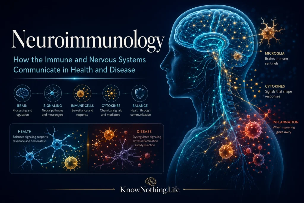
Neuroimmunology is the study of communication between the nervous system and the immune system. It examines how immune cells, antibodies, cytokines, glial cells, blood vessels, and lymphatic structures influence the brain, spinal cord, and peripheral nerves. The field also investigates how neural activity regulates immunity throughout the body. What was once divided into two largely separate branches of medicine is now understood as an interconnected system in which immune signals can alter neural function and neural circuits can shape inflammation, infection responses, metabolism, and tissue repair.
These interactions are essential for normal health. Immune-related molecules help guide brain development, remove damaged cells, remodel neural connections, and defend nervous tissue from pathogens. Problems arise when protective responses become excessive, misdirected, prolonged, or insufficient. Multiple sclerosis, autoimmune encephalitis, neuromyelitis optica spectrum disorder, Guillain–Barré syndrome, myasthenia gravis, and several inflammatory neuropathies are direct examples of neuroimmune disease. Immune activity is also being investigated as a contributor to stroke, traumatic brain injury, chronic pain, epilepsy, Alzheimer’s disease, Parkinson’s disease, and psychiatric symptoms associated with systemic inflammation.
The Brain Is Protected, Not Isolated
The central nervous system was traditionally described as an immune-privileged organ. This concept arose because immune cells enter healthy brain tissue more selectively than they enter many other organs, while transplanted tissue and inflammatory reactions can behave differently within the brain. The blood–brain barrier reinforces this protection through specialized endothelial cells connected by tight junctions. Pericytes, astrocytic end-feet, basement membranes, and immune signaling molecules help determine which substances and cells can pass between circulating blood and nervous tissue. The barrier is not an impermeable wall; it is a regulated interface that transports nutrients, removes waste, and communicates information about infection and systemic inflammation.
The discovery of meningeal lymphatic vessels further changed the idea of complete immune separation. In 2015, Antoine Louveau and colleagues identified functional lymphatic vessels lining the dural sinuses in mice. The vessels carried fluid and immune cells from cerebrospinal fluid toward deep cervical lymph nodes. Later imaging research identified comparable lymphatic structures along dural venous sinuses and cranial nerves in humans. These findings showed that the tissues surrounding the brain participate in immune surveillance and drainage, although many questions remain about how these pathways operate in human disease.
Microglia and the Brain’s Resident Immune System
Microglia are the principal resident immune cells within the brain and spinal cord. Unlike most immune cells, they do not normally arise from adult bone marrow. In a landmark 2010 Science study, Florent Ginhoux and colleagues used fate-mapping experiments to show that adult mouse microglia originate from primitive macrophage precursors that enter the developing brain early in embryonic life. Once established, these cells largely maintain their population through local self-renewal. Microglia continually survey their environment, responding to damaged cells, infectious agents, altered neural activity, and changes in the chemical conditions surrounding neurons.
Microglia do more than defend against disease. During development, they help refine neural circuits by removing unnecessary synapses. Beth Stevens and colleagues demonstrated in 2007 that proteins from the classical complement system, better known for marking pathogens and damaged cells, also participate in normal synapse elimination. Complement proteins can tag selected neural connections, allowing microglia to identify and engulf them. This mechanism is valuable during development, but its inappropriate reactivation may contribute to disease. In Alzheimer’s mouse models, Soyon Hong and colleagues found that blocking complement components or microglial complement receptors reduced early synapse loss.
Astrocytes, Barriers, and Inflammatory Signaling
Astrocytes are star-shaped glial cells that support neurons, regulate neurotransmitters, maintain metabolic conditions, and contribute to the blood–brain barrier. During infection or injury, astrocytes can release cytokines and chemokines that recruit or regulate immune cells. They can also form protective boundaries around damaged tissue. These responses may limit the spread of inflammation, but prolonged or dysregulated astrocyte activity can interfere with neural repair and sustain inflammatory signaling.
The behavior of glial cells cannot be reduced to simple labels such as “protective” or “harmful.” Their effects depend on timing, location, disease stage, and surrounding molecular signals. Keisuke Haruwaka and colleagues showed that microglia initially accumulated around cerebral blood vessels during systemic inflammation and helped protect barrier integrity. Under prolonged inflammatory conditions, however, activated microglia damaged astrocytic end-feet and increased blood–brain barrier permeability. The same immune population can therefore defend nervous tissue during one phase of a response and contribute to injury during another.
Multiple Sclerosis and Misguided Immunity
Multiple sclerosis is among the best-studied neuroimmunological disorders. It involves inflammatory attacks against myelin, the insulating material surrounding many central nervous system axons. Demyelination disrupts signal transmission and may be followed by axonal injury and neurodegeneration. T cells were once considered the dominant drivers of the disease, but clinical trials targeting B cells demonstrated that B-cell functions are also central. B cells may present antigens to T cells, release inflammatory cytokines, produce antibodies, and organize immune activity within the cerebrospinal fluid and meninges.
In a 2008 placebo-controlled trial, Stephen Hauser and colleagues found that the B-cell-depleting antibody rituximab reduced inflammatory brain lesions and clinical relapses in relapsing–remitting multiple sclerosis. Two later phase 3 trials showed that ocrelizumab produced lower relapse rates and less MRI disease activity than interferon beta-1a. The ORATORIO trial also found that ocrelizumab reduced clinical and imaging progression in primary progressive multiple sclerosis. These results changed both treatment and scientific understanding by demonstrating that selectively modifying one immune-cell population can meaningfully alter a complex neurological disease.
Neuromyelitis optica spectrum disorder illustrates a related but biologically distinct form of autoimmunity. Many affected patients produce antibodies against aquaporin-4, a water-channel protein concentrated on astrocytes. Attacks often involve the optic nerves and spinal cord and can cause severe vision loss, weakness, sensory changes, or autonomic dysfunction. Treatment increasingly targets specific immune pathways. In a randomized trial, satralizumab, an antibody directed against the interleukin-6 receptor, reduced relapse risk when added to existing immunosuppressive treatment, demonstrating the value of matching therapy to a defined immune mechanism.
Autoimmune Encephalitis and Brain-Targeted Antibodies
Autoimmune encephalitis occurs when antibodies or immune cells interfere with proteins required for normal brain function. Symptoms may include memory loss, seizures, confusion, abnormal movements, sleep disruption, speech difficulties, psychosis, or changes in consciousness. Some forms were historically mistaken for primary psychiatric illness, viral encephalitis, or unexplained epilepsy. The identification of antibodies against neuronal surface proteins has allowed clinicians to recognize syndromes that may improve with immunotherapy and, when present, treatment of an associated tumor.
Anti-NMDA receptor encephalitis is a major example. In a 2008 study, Josep Dalmau and colleagues described the clinical features of the disorder and demonstrated that patients’ antibodies reduced the number of NMDA receptors on neuronal surfaces. The effect was reversible after the antibodies were removed, helping explain how profound psychiatric and neurological symptoms could occur without widespread permanent neuronal destruction. Patients who received early tumor treatment and immunotherapy generally had better outcomes and fewer relapses, establishing a direct link between antibody biology, clinical presentation, and treatment response.
Neuroinflammation in Degeneration, Infection, and Injury
Neuroinflammation is also present in diseases not traditionally classified as autoimmune. In Alzheimer’s disease, amyloid accumulation, damaged synapses, complement proteins, astrocytes, and microglia interact over many years. Microglia may initially remove debris and restrict injury, yet chronic activation can alter synaptic function and release inflammatory mediators. The 2016 study by Hong and colleagues showed in mouse models that complement-dependent microglial activity contributed to synaptic loss before extensive plaque formation. These findings support an immune contribution to degeneration but do not mean Alzheimer’s disease is caused by inflammation alone.
Infection and injury reveal the same balance between protection and damage. Immune responses are needed to control viruses, bacteria, and damaged tissue, but prolonged complement activation and microglial phagocytosis can affect healthy neural connections. Research in viral models has linked a complement–microglial pathway to memory impairment and synapse loss. Experimental traumatic brain injury has also been shown to disrupt meningeal lymphatic drainage, intensify neuroinflammation, and worsen cognitive outcomes in animals. These observations are helping researchers understand why neurological symptoms may persist after the initial infectious or traumatic event has resolved.
Diagnosis, Treatment, and the Future of Neuroimmunology
Diagnosing a neuroimmune disorder may require neurological examination, MRI, cerebrospinal fluid analysis, electrophysiology, antibody testing, and evaluation for infection or cancer. Results must be interpreted in the context of the clinical syndrome because antibodies detected in blood are not always evidence that they are causing disease. Treatment may involve corticosteroids, intravenous immunoglobulin, plasma exchange, B-cell-depleting antibodies, complement inhibitors, cytokine-targeting drugs, or longer-term immunosuppression. The challenge is to stop destructive immunity without leaving the patient dangerously vulnerable to infection.
The future of neuroimmunology lies in greater precision. Single-cell sequencing is revealing distinct microglial, astrocytic, lymphocyte, and macrophage states that were previously grouped together. Improved imaging may allow clinicians to observe neuroinflammation and lymphatic function more directly, while biomarkers could distinguish active immune injury from irreversible neurological damage. Therapies may eventually suppress harmful immune programs while preserving defense, repair, and normal synaptic maintenance.
Neuroimmunology has overturned the belief that the brain operates separately from the immune system. Nervous tissue is protected by specialized barriers, but it remains in constant communication with immune cells, lymphatic structures, circulating molecules, and the rest of the body. That communication supports development, defense, and repair, yet it can also produce severe disease when directed against myelin, astrocytes, receptors, synapses, or neurons. Understanding this balance is creating more accurate diagnoses and treatments while revealing that brain health is inseparable from immune health.


