
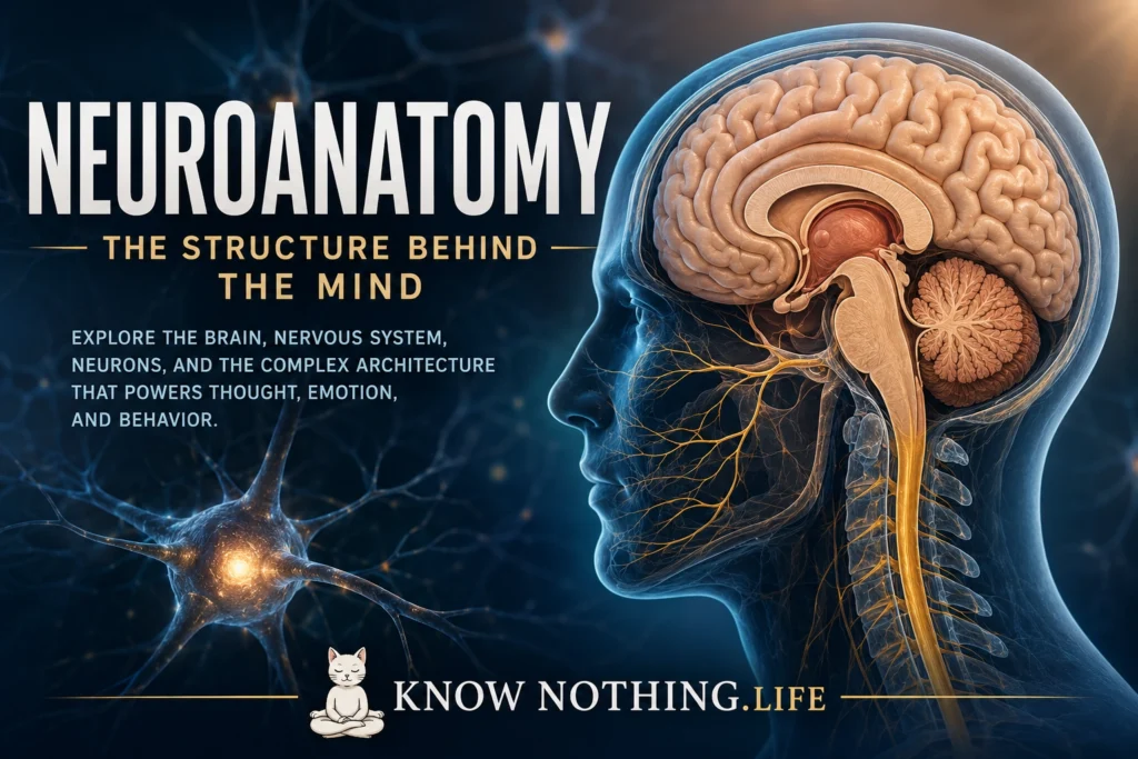
Neuroanatomy is the study of the structure of the nervous system. It asks how the brain, spinal cord, nerves, neurons, glial cells, and sensory pathways are organized, connected, and specialized. If psychology studies thought, emotion, perception, memory, and behavior, neuroanatomy studies the biological architecture that makes those functions possible. It is the map beneath the mind: the physical arrangement of tissue, pathways, regions, and networks through which human experience is generated and regulated.
The importance of neuroanatomy is that structure and function are deeply connected. A stroke in one part of the brain can impair speech. Damage to another area can affect movement, memory, emotion, vision, balance, or personality. The nervous system is not a shapeless mass of tissue. It is highly organized, with specialized regions communicating through electrical and chemical signals. To understand neuroanatomy is to understand how the body senses the world, how the brain interprets it, how movement is controlled, and how consciousness depends on living tissue.
The Nervous System: Central and Peripheral
The nervous system is usually divided into the central nervous system and the peripheral nervous system. The central nervous system includes the brain and spinal cord. It receives information, processes it, coordinates responses, and supports higher functions such as thought, language, memory, emotion, and awareness. The peripheral nervous system includes the nerves outside the brain and spinal cord. These nerves carry sensory information into the central nervous system and motor commands back out to muscles and organs.
The peripheral nervous system is further divided into the somatic and autonomic systems. The somatic nervous system controls voluntary movement and carries sensory information from skin, muscles, and joints. The autonomic nervous system regulates involuntary functions such as heart rate, digestion, breathing patterns, sweating, pupil dilation, and blood pressure. It includes the sympathetic system, associated with arousal and fight-or-flight responses, and the parasympathetic system, associated with rest, digestion, and recovery. Together, these systems allow the nervous system to govern both conscious action and automatic survival.
Neurons and Glial Cells
The basic signaling cells of the nervous system are neurons. A neuron typically has a cell body, dendrites, and an axon. Dendrites receive signals from other neurons, while the axon sends signals outward. At the end of the axon, synapses allow neurons to communicate through neurotransmitters such as glutamate, GABA, dopamine, serotonin, acetylcholine, and norepinephrine. This communication is the foundation of sensation, movement, learning, emotion, and cognition.
Neurons, however, are not alone. Glial cells are essential to nervous system function. Astrocytes help regulate the chemical environment around neurons and support synaptic function. Oligodendrocytes in the central nervous system and Schwann cells in the peripheral nervous system produce myelin, a fatty insulating layer that helps electrical signals travel quickly along axons. Microglia serve immune and cleanup roles in the brain. For much of history, glia were treated as mere support cells, but modern neuroscience shows they are active participants in development, repair, metabolism, and communication.
Gray Matter and White Matter
The nervous system contains gray matter and white matter. Gray matter is rich in neuron cell bodies, dendrites, and synapses. It is where much information processing occurs. The cerebral cortex, the outer layer of the brain, is gray matter. Deep brain nuclei, such as the basal ganglia and thalamus, are also gray matter structures. White matter consists largely of myelinated axons that connect different regions. It appears white because of the fatty myelin surrounding many nerve fibers.
This distinction is important because the brain works through both local processing and long-distance communication. Gray matter regions perform specialized computations, while white matter tracts connect those regions into networks. The corpus callosum, for example, is a major white matter pathway connecting the left and right cerebral hemispheres. Damage to white matter can disrupt communication even when individual brain regions remain intact. In this sense, neuroanatomy is not only about locations; it is about connections.
The Cerebral Cortex
The cerebral cortex is the wrinkled outer surface of the brain and is especially important for perception, voluntary movement, reasoning, language, attention, memory, and consciousness. Its folds, called gyri and sulci, increase surface area and allow more cortical tissue to fit inside the skull. The cortex is divided into two hemispheres, left and right, and each hemisphere is organized into major lobes: frontal, parietal, temporal, and occipital. Some classifications also include the insula and limbic cortex.
The frontal lobe is involved in planning, decision-making, voluntary movement, attention, impulse control, and aspects of personality. The parietal lobe helps process touch, spatial awareness, body position, and sensory integration. The temporal lobe is important for hearing, language comprehension, memory, and object recognition. The occipital lobe processes visual information. These divisions are useful, but the brain rarely works in isolated boxes. Most complex functions depend on networks spanning multiple areas.
The Limbic System and Emotion
The limbic system is a group of structures often associated with emotion, memory, motivation, and survival behavior. Important limbic structures include the amygdala, hippocampus, hypothalamus, cingulate cortex, and parts of the thalamus. The amygdala is involved in emotional salience, especially fear, threat detection, and learning from emotionally important events. The hippocampus is essential for forming new declarative memories and for spatial navigation. Damage to the hippocampus can severely impair the ability to create new long-term memories.
The hypothalamus is small but powerful. It regulates hunger, thirst, temperature, sleep-wake rhythms, hormonal activity, sexual behavior, and stress responses. It links the nervous system with the endocrine system through the pituitary gland. This connection shows that neuroanatomy is not limited to abstract thought. The brain is deeply embodied. Emotion, memory, hormones, and bodily regulation are inseparable from nervous system structure.
The Basal Ganglia and Movement
The basal ganglia are deep brain structures involved in movement, habit formation, reward, and action selection. They include the caudate nucleus, putamen, globus pallidus, substantia nigra, and related structures. These circuits help initiate desired movements and inhibit unwanted movements. They are also involved in procedural learning, such as habits and skilled actions.
Diseases of the basal ganglia show their importance. Parkinson’s disease involves degeneration of dopamine-producing neurons in the substantia nigra, leading to tremor, rigidity, slowness of movement, and postural difficulties. Huntington’s disease affects basal ganglia circuits and produces involuntary movements, cognitive changes, and psychiatric symptoms. These disorders reveal that movement is not simply commanded by the motor cortex; it depends on deep regulatory loops that help select, refine, and coordinate action.
The Thalamus and Brainstem
The thalamus is often described as a relay station, but that description is too simple. It helps route sensory and motor information to the cortex and participates in attention, consciousness, and sleep-wake regulation. Nearly all sensory information, except smell, passes through thalamic circuits before reaching the cortex. The thalamus does not merely pass information along; it helps regulate what becomes available to conscious processing.
The brainstem connects the brain to the spinal cord and includes the midbrain, pons, and medulla. It controls many basic life functions, including breathing, heart rate, arousal, swallowing, and reflexes. It also contains pathways that carry information between the brain and body. The reticular formation, a network within the brainstem, plays an important role in wakefulness and alertness. Damage to the brainstem can be life-threatening because it supports the most fundamental conditions of survival.
The Cerebellum
The cerebellum, located at the back of the brain beneath the cerebral hemispheres, is traditionally associated with balance, coordination, timing, and motor learning. It helps movements become smooth and accurate. A person with cerebellar damage may have poor coordination, tremor during movement, difficulty with balance, and problems adapting motor actions.
Modern neuroscience also connects the cerebellum to cognition, language, emotion, and prediction. Its dense circuitry appears to help the brain compare intended actions with actual outcomes, allowing correction and learning. This predictive function may extend beyond movement into thought and behavior. The cerebellum reminds us that structures once considered purely motor may also contribute to higher mental processes.
The Spinal Cord and Nerves
The spinal cord is the major communication highway between the brain and body. It carries sensory information upward and motor commands downward. It is organized into cervical, thoracic, lumbar, sacral, and coccygeal regions. Spinal nerves branch from the cord and connect with muscles, skin, and organs. Reflexes can be coordinated at the spinal level without requiring immediate conscious control from the brain. For example, pulling the hand away from a painful stimulus can occur rapidly through spinal reflex circuits.
The spinal cord’s organization is clinically important. Injury at different levels produces different patterns of impairment. Damage in the cervical region may affect arms, legs, and breathing, while lower injuries may affect legs and pelvic function. Neuroanatomy therefore has direct medical relevance. It helps clinicians localize damage, understand symptoms, and predict functional outcomes.
Cranial Nerves and Sensory Systems
The brain has twelve pairs of cranial nerves that support smell, vision, eye movement, facial sensation, facial expression, hearing, balance, taste, swallowing, speech, and autonomic regulation. These nerves include the optic nerve for vision, the trigeminal nerve for facial sensation and chewing, the facial nerve for facial expression, the vestibulocochlear nerve for hearing and balance, and the vagus nerve, which influences many organs in the chest and abdomen.
Sensory systems are organized through specialized pathways. Vision travels from the retina through the optic nerve, optic chiasm, thalamus, and visual cortex. Hearing travels from the inner ear through brainstem nuclei to auditory cortex. Touch, pain, temperature, and body position travel through spinal pathways to the thalamus and somatosensory cortex. Each sensory system transforms physical energy into neural signals, allowing the brain to construct a usable world.
Final Thoughts
Neuroanatomy is the study of the nervous system’s architecture, but it is also a foundation for understanding human experience. The frontal lobes help organize action and judgment. The temporal lobes support memory and meaning. The occipital lobes process vision. The limbic system links emotion and memory. The basal ganglia shape movement and habit. The brainstem sustains life. The cerebellum refines coordination and prediction. The spinal cord connects brain and body. Neurons and glia build the microscopic machinery beneath it all.
The power of neuroanatomy lies in showing that mind and body are not separate worlds. Perception, emotion, thought, movement, memory, and consciousness depend on living structure. Every human experience has a biological pathway, but those pathways form an astonishingly complex system. To study neuroanatomy is to study how matter becomes sensation, how cells become networks, how networks become behavior, and how the nervous system gives rise to the fragile, embodied life of the mind.


